
Fig. 3
- ID
- ZDB-IMAGE-250421-75
- Publication
- Huybrechts et al., 2025 - Loss of the Ubiquitin-Associated Domain of sqstm1/p62 in Zebrafish Causes a Phenotype Resembling Paget's Disease of Bone
- All Figures
- Figures for Huybrechts et al., 2025
|
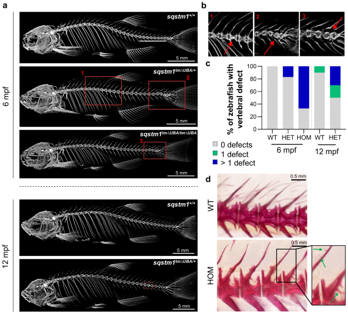
Fig. 3 Vertebral columns of sqstm1tmΔUBA zebrafish are characterized by structural defects and ectopic mineralization. a Representative µCT images at 6 and 12 mpf. Vertebral malformations, including fused vertebrae, are evident in carriers of one or two mutant sqstm1 alleles, mainly in the precaudal to caudal vertebral junction and/or the tail region (red squares). b Close-up of the red squares shown in (a), labeled by 1–3. c Quantification of animals with certain number of vertebral defects. For 6 mpf, n = 4 (WT) or 6 (HET/HOM) zebrafish/genotype and for 12 mpf, n = 10 zebrafish/genotype. d Alizarin Red staining of a wild-type and homozygous zebrafish, including a magnification box. Ectopic mineralization and osteophytes affecting the whole vertebral column are shown by green arrows. WT wild type (sqstm1+/+), HET heterozygous mutant (sqstm1tmΔUBA/+), HOM homozygous mutant (sqstm1.tmΔUBA/tmΔUBA)