IMAGE

Fig. 4

ID
ZDB-IMAGE-250418-6
Source
Figures for Murcott et al., 2025
Image
Figure Caption

Fig. 4

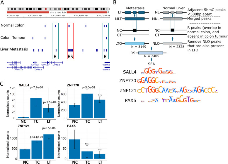
In liver metastasis, 5hmC peaks that were initially present in normal colon are recovered. A IGV screenshot with uploaded tracks of 5hmC consensus peaks for normal colon, colon tumour and liver metastasis. Boxes above R highlight a 5hmC peak that has ‘recovered’ since it is present in normal colon, absent in colon tumours and reappears in liver metastasis. Boxes above RS highlight a 5hmC peak that has ‘recovered and spread’. Thus, the peak is present in normal colon, lost in tumour colons and reappears in metastasis, but also spreads to adjacent sites. B The strategy for identifying RS peaks and the consensus binding sites for SALL4, ZNF770, ZNF121 and PAX5 identified after SEA. Abbreviations: MLT and MNL are Merged liver tumour (LT) and normal liver (NL) peaks respectively. LTO and NLO are LT or NL peaks that Overlap normal colon. C Normalised RNAseq counts (data used [28]) for SALL4, ZNF770, ZNF121 and PAX5 in normal colon, colon tumours and liver metastasis (NC, CT and LM). Wald test, Benjamini-Hochberg adjusted. Error bars represent ± SEM. Supporting PCR data values in Additional file 6

Acknowledgments
This image is the copyrighted work of the attributed author or publisher, and ZFIN has permission only to display this image to its users. Additional permissions should be obtained from the applicable author or publisher of the image. Full text @ BMC Biol.