IMAGE

Fig. 5.

ID
ZDB-IMAGE-250417-96
Source
Figures for Zhao et al., 2025
Image
Figure Caption

Fig. 5.

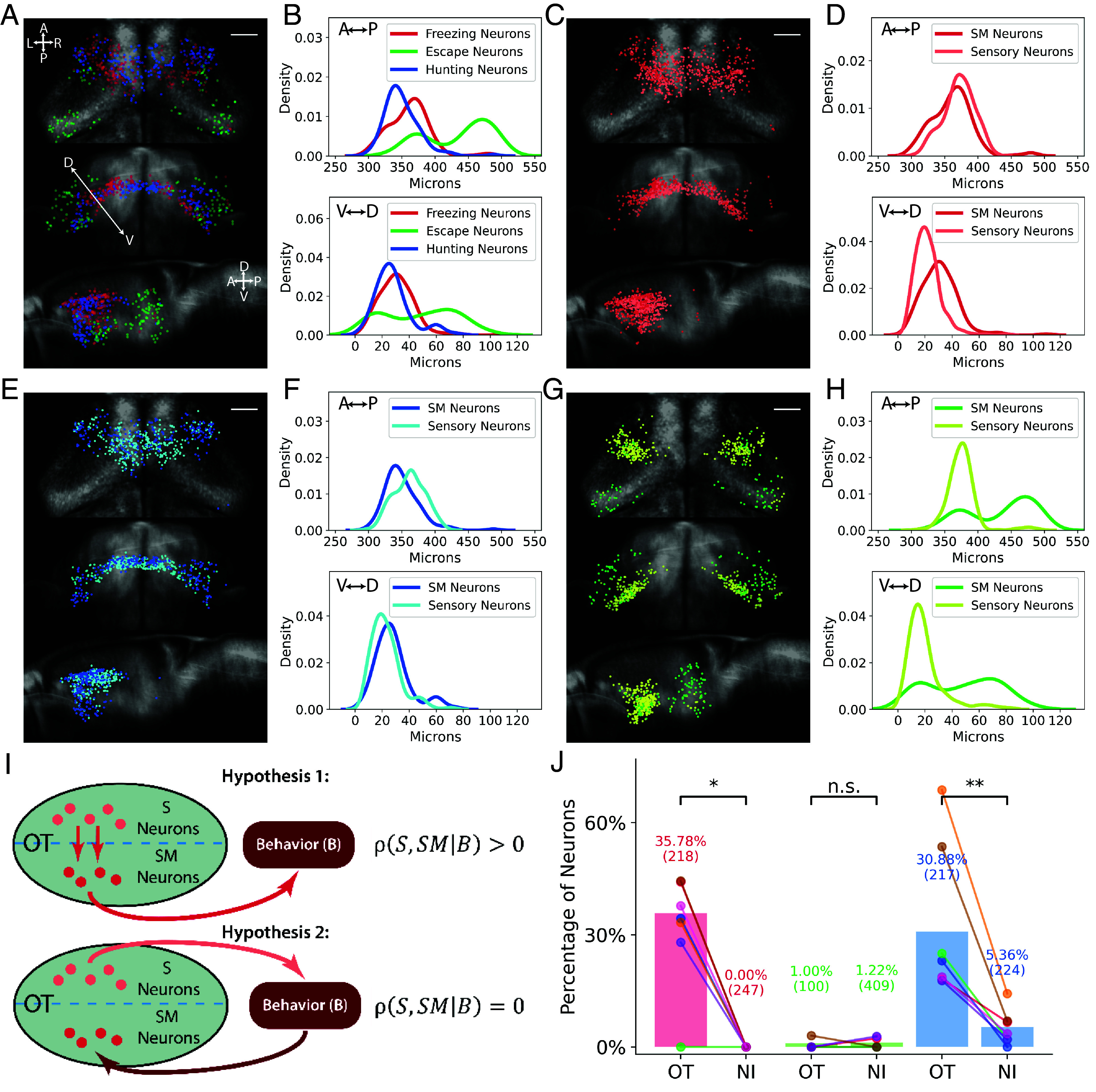
Sensory and sensorimotor neurons in the tectum. (A) Locations of the three types of SM neurons in the tectum. (Scale bar, 50 µm.) (B) Locations of SM neurons on the anterior/posterior and dorsal/ventral axes. (C and D) Locations of sweep sensory neurons (light red) and freezing SM neurons (dark red) in the tectum and their locations on the anterior/posterior and dorsal/ventral axes. (E and F) Locations of prey sensory neurons (light blue) and hunting SM neurons (dark blue) in the tectum and on the anterior/posterior and dorsal/ventral axes. (G and H) Locations of looming sensory neurons (light green) and escape SM neurons (green) in the tectum and their locations on the anterior/posterior and dorsal/ventral axes. (I) Hypotheses for the tectum S and SM connectivity in the sensorimotor circuit. (J) Percentage of SM neurons that were significantly correlated with the tectal sensory neurons. Colored dots represent values for individual fish. Parenthesis: total number of SM neurons of that type across all fish. NI = Percentage of SM neurons in the NI that were significantly correlated with tectal sensory neurons. Percentage-by-chance was 0% for both tectal and NI SM neurons (Methods). *, P < 0.05. **, P < 0.01, one-sided Wilcoxon signed-rank test. The P-values were 0.011 for freezing, 0.787 for escape, and 0.007 for hunting.

Acknowledgments
This image is the copyrighted work of the attributed author or publisher, and ZFIN has permission only to display this image to its users. Additional permissions should be obtained from the applicable author or publisher of the image. Full text @ Proc. Natl. Acad. Sci. USA