
|
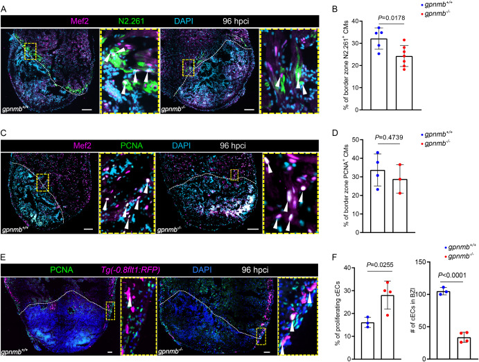
Fig. 4 gpnmb mutants exhibit reduced cardiomyocyte dedifferentiation and reduced coronary endothelial cell numbers after cardiac cryoinjury in zebrafish. A. Immunostaining for Mef2 (cardiomyocyte nuclei, green), N2.261 (embryonic myosin heavy chain, magenta) with DAPI (DNA marker, cyan) counterstaining on sections of gpnmb+/+ and gpnmb−/− cryoinjured ventricles at 96 hpci. B. Quantification of dedifferentiating cardiomyocytes in border zone areas (100 μm) at 96 hpci. C. Immunostaining for Mef2 (cardiomyocyte nuclei, green), PCNA (proliferation marker, magenta) with DAPI (DNA marker, cyan) counterstaining on sections of gpnmb+/+ and gpnmb−/− cryoinjured ventricles at 96 hpci. D. Quantification of proliferating cardiomyocytes in border zone areas (100 μm) at 96 hpci. E. Immunostaining for PCNA (proliferation marker, green), RFP (coronary endothelial cells, magenta) with DAPI (DNA marker, blue) counterstaining on sections of Tg(-0.8flt1:RFP); gpnmb+/+ and Tg(-0.8flt1:RFP); gpnmb−/− cryoinjured ventricles at 96 hpci. F. Quantification of proliferating and total number of coronary endothelial cells in the BZI tissue (200 μm) at 96 hpci. White dotted lines delineate the injured area; white arrowheads point to dedifferentiating cardiomyocytes (A), proliferating cardiomyocytes (C), and proliferating coronary endothelial cells (E). Statistical test: Student's t-test (B, D, F). Scale bars: 100 μm. (For interpretation of the references to color in this figure legend, the reader is referred to the Web version of this article.)
Reprinted from Developmental Biology, 521, Gupta, S., Bajwa, G.K., El-Sammak, H., Mattonet, K., Günther, S., Looso, M., Stainier, D.Y.R., Marín-Juez, R., The transmembrane glycoprotein Gpnmb is required for the immune and fibrotic responses during zebrafish heart regeneration, 153-162, Copyright (2025) with permission from Elsevier. Full text @ Dev. Biol.