
Image
|
Figure Caption
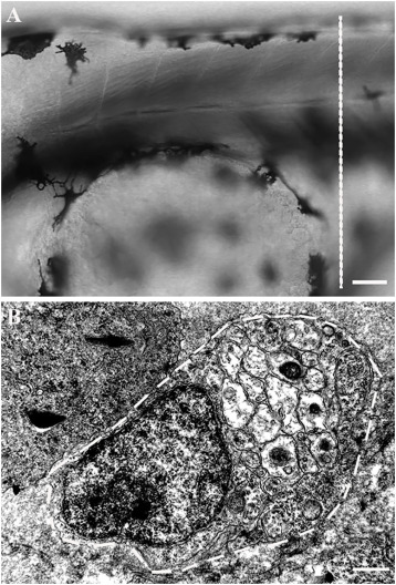
Fig. S1 Radial sorting along the anterior-posterior (AP) axis of the PLLn. (A) Lateral view of a 48 hpf embryo. The dotted line represents the AP position of the cross-section analysis by TEM. Anterior is to the left and dorsal to the top. Scale bar = 50 μm. (B) TEM of a cross-section of the PLLn of a WT embryo at 48 hpf. The PLLn is delineated in white dotted lines. Only 2 axons out of 94 (≈2 %) were radially sorted at 48 hpf at this AP position (5 nerves, n = 5 embryos). Scale bar = 0.5 μm.
Acknowledgments
This image is the copyrighted work of the attributed author or publisher, and
ZFIN has permission only to display this image to its users.
Additional permissions should be obtained from the applicable author or publisher of the image.
Full text @ Cells Dev