IMAGE

Figure 4

ID
ZDB-IMAGE-250328-9
Source
Figures for Besio et al., 2025
Image
Figure Caption

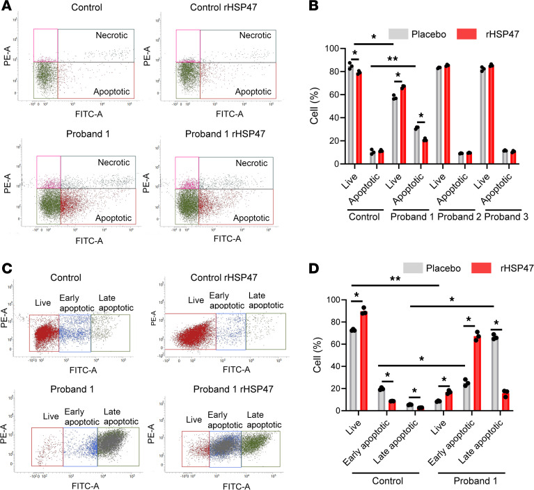
Figure 4 rHSP47 modulates cell apoptosis.

(A) FACS analysis detection of apoptotic cells in OI probands and control fibroblasts treated with rHSP47 or with placebo following annexin V (FITC) and propidium iodide (PI) staining. The fraction of apoptotic events in the cells is shown in representative plots and (B) quantified in the histogram. Biological replicates (n = 3) were performed. Error bars indicate SD. (C) FACS analysis of apoptotic cells in OI proband 1 and control fibroblasts treated with rHSP47 or with placebo following a high-stress condition induced by culturing the cells for 7 days without media change. The fraction of apoptotic events in the cells is shown in representative plots and (D) quantified in the histogram. Both early and late apoptotic cells were quantified. Biological replicates (n = 3) were performed. Error bars indicate SD. Mann-Whitney Wilcoxon test was performed. Statistical analyses details are reported in Supplemental Table 2. *P < 0.05; **P ≤ 0.01.

Acknowledgments
This image is the copyrighted work of the attributed author or publisher, and ZFIN has permission only to display this image to its users. Additional permissions should be obtained from the applicable author or publisher of the image. Full text @ JCI Insight