IMAGE

Figure 3

ID
ZDB-IMAGE-250328-8
Source
Figures for Besio et al., 2025
Image
Figure Caption

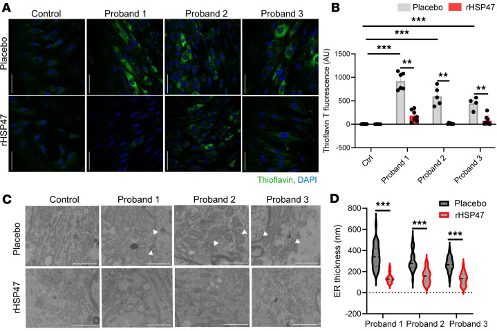
Figure 3 rHSP47 ameliorates cellular homeostasis.

ER proteostasis was evaluated by thioflavin T (ThT) labeling of protein aggregates in osteogenesis imperfecta (OI) proband and control fibroblasts treated for 16 hours with 0.5 μM rHSP47 or with placebo. (A) Representative immunofluorescence images and (B) ThT quantification (aligned rank ANOVA [ARA] test, FARA = 86.82) is shown (P < 0.001 by overall test; asterisks indicate post hoc test results). Biological replicates (n = 3) were performed. For each biological replicate, the signal was quantified on 7 images (original magnification, ×40) for each genotype/condition (number of cells >70). Error bars indicate SD. Scale bars: 20 μm. (C) Representative transmission electron microscopy images of OI probands and control fibroblasts treated for 16 hours with 0.5 μM rHSP47 or with placebo (n = 3). Arrowheads show ER enlargement. Scale bars: 2 μm. (D) ER enlargement was quantified for 30 proband cells treated with rHSP47 or with placebo. Mann-Whitney Wilcoxon test, MW = 6.35 (P < 0.001 by overall test; asterisks indicate post hoc test results). Statistical analyses details are reported in Supplemental Table 2. **P ≤ 0.01; ***P ≤ 0.001.

Acknowledgments
This image is the copyrighted work of the attributed author or publisher, and ZFIN has permission only to display this image to its users. Additional permissions should be obtained from the applicable author or publisher of the image. Full text @ JCI Insight