IMAGE

Fig 2

ID
ZDB-IMAGE-250321-11
Source
Figures for Nonarath et al., 2025
Image
Figure Caption

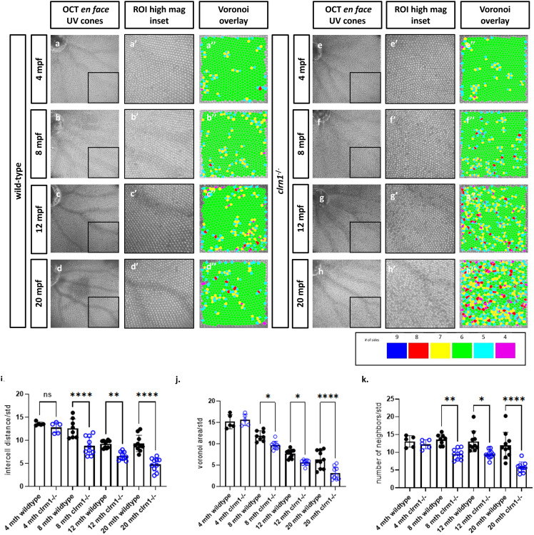
Fig 2 clrn1-/- zebrafish present with an altered UV cone mosaic beginning at 8 mpf.

En face images of the UV cone mosaic were segmented from OCT scans, a higher magnification ventro-temporal region of interest (ROI) was selected (a’-h’) and processed for Voronoi overlay analysis (a”-h”) in wild-type at (a) 4, (b) 8, (c) 12, and (d) 24 mpf, as well as clrn1-/- zebrafish at (e) 4, (f) 8, (g) 12, and (h) 24 mpf. Quantification of the UV cone mosaic regularity revealed statistically significant differences in (i) intercell distance regularity, (j) number of neighbors regularity and, (k) the Voronoi area regularity at 8 mpf in clrn1-/- zebrafish compared to wild-type. (*p<0.05, **p<0.01, ****p<0.001; Two-way ANOVA).

Acknowledgments
This image is the copyrighted work of the attributed author or publisher, and ZFIN has permission only to display this image to its users. Additional permissions should be obtained from the applicable author or publisher of the image. Full text @ PLoS Genet.