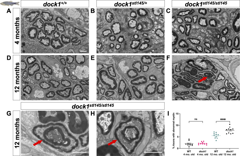
Age-dependent myelin maintenance defects are present in dock1 MUTs. (A–C) TEM of cross-sections of ZMBs from 4-mo-old WT dock1+/+, HET dock1stl145/+, and homozygous dock1stl145/stl145 MUT zebrafish. (D–F) TEM micrographs of ZMBs from 12-mo-old WT dock1+/+, HET dock1stl145/+, and homozygous dock1stl145/stl145 MUT zebrafish, with homozygous MUTs exhibiting myelin outfoldings (red arrow). (G and H) Higher-magnification TEM micrographs of ZMBs from 12-mo-old homozygous dock1stl145/stl145 MUTs showing abnormally myelinated axons (red arrows), features rarely seen in WT dock1+/+ or HET dock1stl145/+ animals. (I) Quantification of the percent of axons with abnormal myelin profiles, observed by TEM, in WT dock1+/+ versus homozygous dock1stl145/stl145 MUTs at 4 and 12 mo old, n = 10 (WT dock1+/+ 4 mo old), 10 (dock1stl145/stl145 MUT 4 mo old), 10 (WT dock1+/+ 12 mo old), and 10 (dock1stl145/stl145 MUT 12 mo old). Here, and in all figures, the X symbol in the graph denotes a data point corresponding to the representative image shown. (A–H) Scale bar = 1 µm. (I) Two-way ANOVA with Tukey’s multiple comparisons test. ***P < 0.001; ns, not significant.
This image is the copyrighted work of the attributed author or publisher, and
ZFIN has permission only to display this image to its users.
Additional permissions should be obtained from the applicable author or publisher of the image.
Full text @ J. Cell Biol.
Your Input Welcome
Thank you for submitting comments. Your input has been emailed to ZFIN curators who may contact you if
additional information is required.
Oops. Something went wrong. Please try again later.