IMAGE

Fig. 6

ID
ZDB-IMAGE-250318-16
Source
Figures for Ahlmann-Eltze et al., 2025
Image
Figure Caption

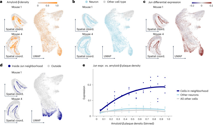
Fig. 6 Analysis of a spatial single-cell experiment.

ad, UMAPs of cells from the hippocampi of four mice and spatial maps for two of them, colored by amyloid-β plaque density (calculated from smoothed and normalized fluorescent images26) (a), whether the cell is a neuron (b), LEMUR-predicted differential expression (log2 fold change) for Jun (c) and whether the cell is inside the differential expression neighborhood for Jun (d). e, Scatterplot of Jun expression as a function of plaque density for cells inside the neighborhood, neurons not in the neighborhood and all other cells. The line is a local regression (LOESS) fit through the pseudobulked values. Coord., coordinate.

Acknowledgments
This image is the copyrighted work of the attributed author or publisher, and ZFIN has permission only to display this image to its users. Additional permissions should be obtained from the applicable author or publisher of the image. Full text @ Nat. Genet.