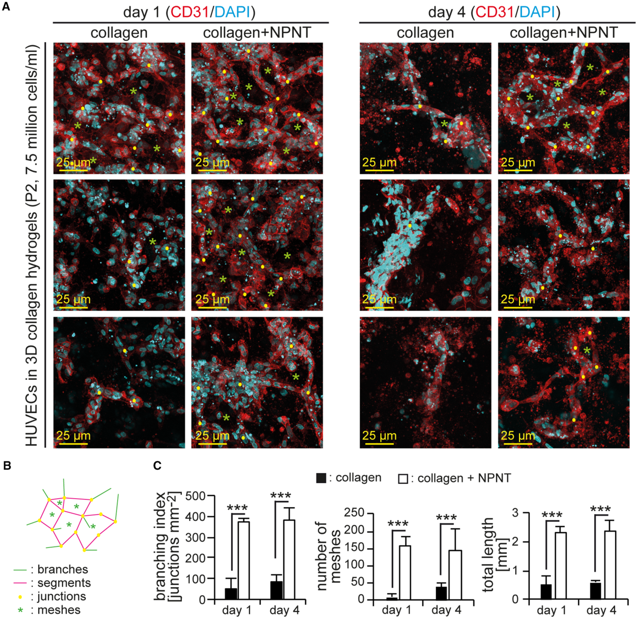
Fig. 10 mr‐NPNT enhances vessel formation in hydrogels. A, Examples of projections of confocal images of HUVECs (passage number 2, P2) cultured in collagen‐I‐based hydrogels stained for endothelial specific marker CD31 and DNA by 4′,6‐diamidino‐2‐phenylindole. Yellow dots: examples of junctions. Green asterisks: examples of meshes. B, Schematic of a vascular network indicating measured parameters. C, Quantitative analysis of (A) (n=3) regarding vasculature complexity based on branching index, number of meshes, and total length (segments + branches). Statistical significance was determined by a 2‐tailed Student's t test. Data are mean±SEM. ***P≤0.001. DAPI indicates 4′,6‐diamidino‐2‐phenylindole; HUVEC, human umbilical vascular endothelial cell; mr‐NPNT, recombinant mouse nephronectin.
Image
Figure Caption
Acknowledgments
This image is the copyrighted work of the attributed author or publisher, and
ZFIN has permission only to display this image to its users.
Additional permissions should be obtained from the applicable author or publisher of the image.
Full text @ J. Am. Heart Assoc.

