
Fig. 8
- ID
- ZDB-IMAGE-250311-82
- Publication
- Patra et al., 2025 - Nephronectin Is Required for Vascularization in Zebrafish and Sufficient to Promote Mammalian Vessel-Like Structures in Hydrogels for Tissue Engineering
- All Figures
- Figures for Patra et al., 2025
|
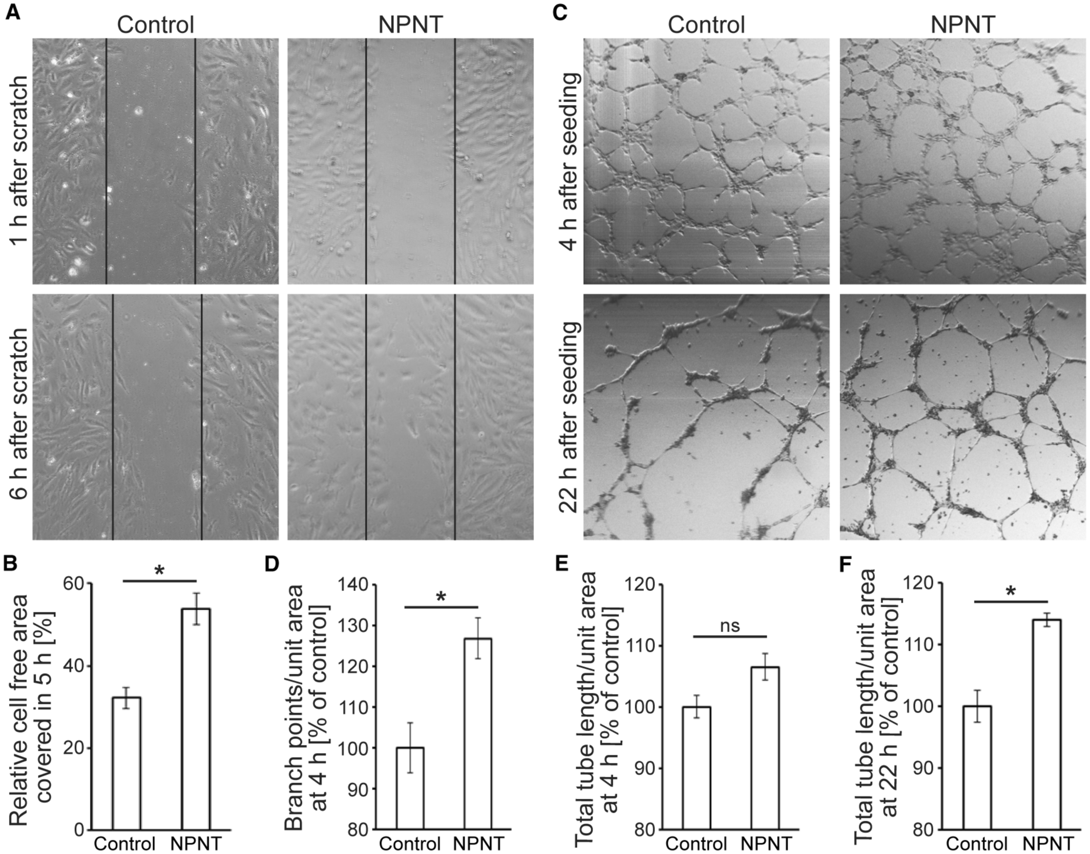
Fig. 8 Nephronectin promotes HUVEC migration, tube formation, and stabilization in vitro. A, Cell migration assay: representative brightfield images of wound assays 1 and 6 hours after scratching. Cells were cultured on gelatin‐ (control) or mr‐NPNT‐coated 24‐well cell culture plates. B, Quantitative analysis of cell migration (n=4). The mean value of the control was considered 100% in each case. C, Tube formation assay: representative brightfield images of HUVECs cultured on Matrigel with BSA (10 μg/mL, control) or mr‐NPNT (10 μg/mL) at 4 and 22 hours after seeding. D–F, Quantitative analysis of tube branching (D), and tube stability (E, F) (n=4). Mean value of the control was considered 100% in each case. Statistical significance was determined by a 2‐tailed Student's t test. Data are mean±SEM. ns: P>0.05. *P≤0.05. HUVEC indicates human umbilical vascular endothelial cell; and mr‐NPNT, recombinant mouse nephronectin.