
|
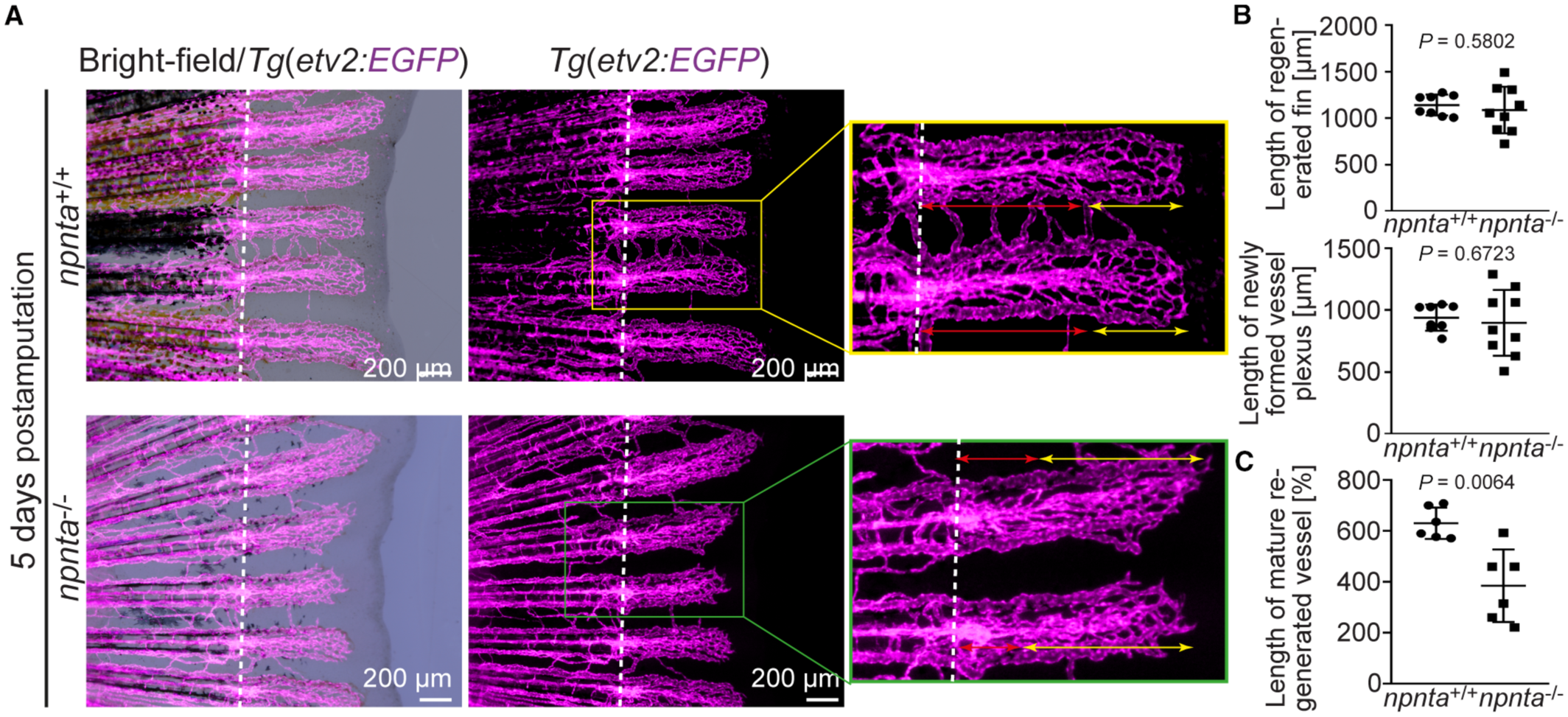
Fig. 7 Adult npnta mutants exhibit decreased vessel pruning during caudal fin regeneration. A, Representative overlays of bright‐field and epifluorescence images as well as epifluorescence images of individual caudal fins of a wild‐type and npnta mutant in an Tg(etv2:EGFP) background at 5 days post amputation revealing decreased vessel maturation (magenta; endothelial cell membrane) during caudal fin regeneration. Double‐sided yellow arrows: length of neovascular plexus. Double‐sided red arrows: length of matured neovessels. Scale bars: 200 μm. B, Quantitative analysis of the length of regenerated fins and the length (μm) of regenerated blood vessels. C, Quantification of the length of the matured blood vessels at the proximal ends of the regenerated fins. Statistical significance was determined by a 2‐tailed Student's t test. Data are mean±SEM. ns: P>0.05, and **P≤0.01.