
Fig. 2
- ID
- ZDB-IMAGE-250304-1
- Publication
- Chen et al., 2025 - NDUFB7 mutations cause brain neuronal defects, lactic acidosis, and mitochondrial dysfunction in humans and zebrafish
- All Figures
- Figures for Chen et al., 2025
|
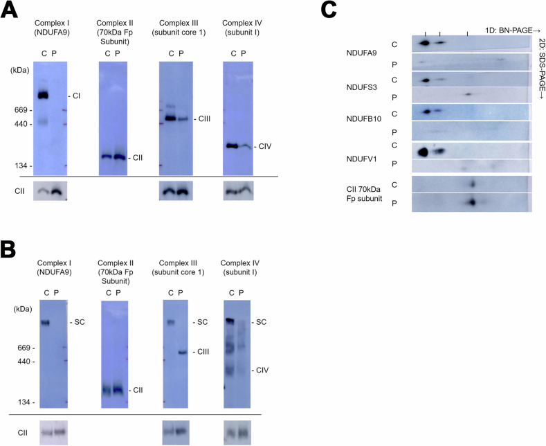
Fig. 2 Mitochondrial complex formation is disrupted in fibroblasts derived from the patient carrying the NDUFB7 mutations.
Using blue native polyacrylamide gel electrophoresis (BN-PAGE), mitochondrial extracts from control (C) and patient (P) skin fibroblasts were separated using Triton