IMAGE

Fig. 4

ID
ZDB-IMAGE-250219-35
Source
Figures for Dharmadhikari et al., 2025
Image
Figure Caption

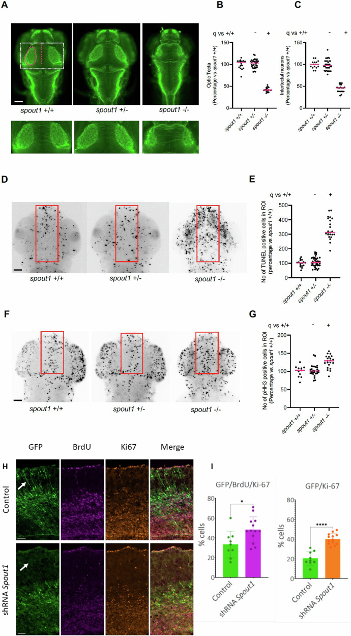
Fig. 4 spout1/cenp-32-/- mutant larvae have CNS defects, apoptosis and altered cell cycle progression.

A Representative dorsal images of wholemount larvae fixed at 3 dpf and immunostained for acetylated tubulin to demarcate axon tracts. We counted commissural axons crossing the midline between the optic tecta (pink dashed line); and the area of optic tecta (pink dashed oval). Dashed white box (upper panel) represents magnified image in lower panel. Scale bar, 100 µM. B, C Quantification of optic tecta size and intertectal neuron number, respectively. D Representative dorsal inverted fluorescent images of zebrafish larvae marked by TUNEL at 2 dpf. The region of interest (ROI) quantified is shown with a red rectangle. Scale bar, 100 µM. E Quantification of TUNEL stained cells as measured in the ROI. F Representative dorsal inverted fluorescent images showing phospho-histone H3 (pHH3) positive cells at 2 dpf. The ROI quantified is shown with a red rectangle. Scale bar, 100 µM. G Quantification of pHH3 positive cells as measured in ROI. Data shown in B, C, E, and G are combined from two biological replicates. Statistical differences were calculated using non-parametric ANOVA with Kruskal-Wallis test followed by Dunn’s multiple comparisons test by controlling False Discovery Rate (original FDR method of Benjamini and Hochberg); ( + ) and (-) indicate significant and non-significant differences, respectively. Median values are shown with pink horizontal lines. See Supplementary Table S2 for exact adjusted q-values and numbers of larvae. H, I Murine cortical progenitor cells electroporated with shRNA against Spout1 less frequently leave mitotic cell cycle both within 24 hours (I, left diagram), and 48 hours (I, right diagram) after electroporation, and also less frequently migrate to the cortical plate (CP) (white arrows) compared to wild type cells (Note: GFP positive neurons in the control CP and lack of them in shRNA electroporated brains). In utero electroporation of targeting constructs into the lateral ventricles was carried out at E13.5. 24 hours later, neocortical progenitors were labeled with BrdU during S phase of the mitotic cycle. Neocortical cells that express both mitotic marker Ki67 and GFP represent a fraction of the cortical cells that still proliferate 48 hours after in utero electroporation, while cells that express BrdU, Ki67 and GFP represent a fraction of cells that proliferate 24 hours after BrdU injection. Statistical differences were calculated using two-sided unpaired t-test (*, P ≤ 0.05; ****, P ≤ 0.0001). I left diagram P = 0.0176. Data are presented as Mean +/- SD. Number of brain samples analyzed: 3 (control), 4 (experiment); number of slices analyzed: 9 (control), 13 (experiment). Scale bar, 50 µM. Source data are provided as a Source Data file. Created in BioRender102.

Acknowledgments
This image is the copyrighted work of the attributed author or publisher, and ZFIN has permission only to display this image to its users. Additional permissions should be obtained from the applicable author or publisher of the image. Full text @ Nat. Commun.