
Image
|
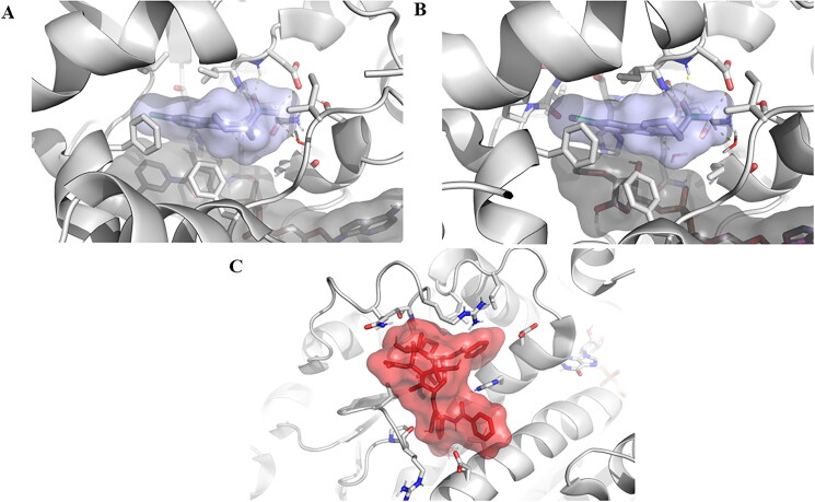
Figure Caption
Figure 8.
Experimentally solved structures of EX527 analogue bound to (a) SIRT1, as revealed by the PDB entry 4I5I. (b) EX527 bound to SIRT3 in the PDB entry 4BVH, (c) Taxol molecule bound to a tubulin unit, as presented by the PDB entry 6WVR. The distinct chemical nature of the ligands, variations in size, and differences in the characteristics of their respective binding pockets render it improbable for them to engage in competition for the same binding site.
Acknowledgments
This image is the copyrighted work of the attributed author or publisher, and
ZFIN has permission only to display this image to its users.
Additional permissions should be obtained from the applicable author or publisher of the image.
Full text @ J Enzyme Inhib Med Chem