IMAGE

Fig. 8

ID
ZDB-IMAGE-250212-42
Source
Figures for Carrara et al., 2025
Image
Figure Caption

Fig. 8

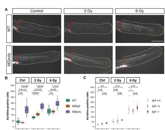
Cell death following irradiation in MMEJ and cNHEJ mutants. 24-hpf embryo from wildtype, MZpolq and MZlig3 and from lig4 heterozygous crosses were exposed to ionizing radiation (0, 2 and 6 Gy) and allowed to develop until 48 hpf. Cell death was then assayed using acridine orange staining (see methods) and quantified in the tail region. (A) Representative images of non-irradiated controls and irradiated with 2 and 6 Gy for wildtype and MZpolq mutant. Note the high number of fluorescent cells in MZpolq in both the control and the irradiated conditions. The red and the yellow lines delimit the area used for quantification. Scale bar: 200 microns. (B-C) Quantification of positive cells in WT, MZpolq, MZlig3 mutants (B) and in lig4 mutant (C). Cell numbers were normalized over the counted area. Note that lig4 mutants were obtained from heterozygous crosses and that embryos were genotyped after staining and scoring. Results from homozygous mutants, heterozygous, and wildtype siblings are presented. p.values from Wilcoxon tests.

Acknowledgments
This image is the copyrighted work of the attributed author or publisher, and ZFIN has permission only to display this image to its users. Additional permissions should be obtained from the applicable author or publisher of the image. Full text @ Sci. Rep.