IMAGE

Fig 5

ID
ZDB-IMAGE-250207-64
Source
Figures for Widziolek et al., 2025
Image
Figure Caption

Fig 5 The role of macrophages and neutrophils in the eradication of Porphyromonas gingivalis (Pg) infection.

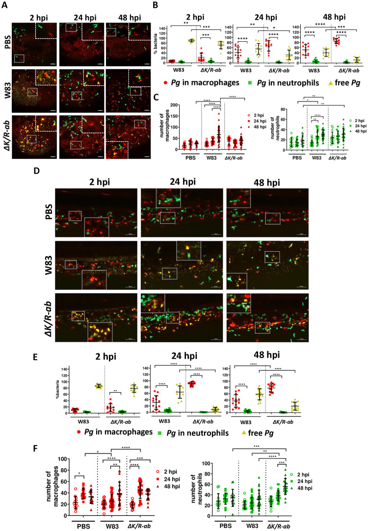
Zebrafish larvae (30 hpf) were infected systemically with AlexaFluor647–SE labelled wild-type Pg W83 or ΔK/R-ab and real-time examination of phagocytosis was performed using transgenic zebrafish larvae Tg(mpeg1:mCherry; red macrophages) and Tg(mpx:EGFP, green neutrophils). PBS was injected as a control. Representative images of the yolk (A) or the tail (D) regions at 2, 24 or 48 hours post-infection (hpi). Scale bar = 50 μm. Quantified percentage of bacteria phagocytosed by macrophages, neutrophils and non-phagocytosed (free) bacteria at 2, 24 and 48 hpi in the yolk (B) and the tail (E) regions. Number of macrophages and neutrophils in the yolk (C) and the tail (F) regions at 2, 24 or 48 hpi. White dashed boxes correspond to zoomed regions of the images. Each dot represents quantified data from a single larva obtained in at least 3 independent experiments. Graphs show means ± SD. Differences between groups were analysed by Two-Way ANOVA.*p≤0.05,**p≤0.01,***p≤0.001,****p≤0.0001.

Acknowledgments
This image is the copyrighted work of the attributed author or publisher, and ZFIN has permission only to display this image to its users. Additional permissions should be obtained from the applicable author or publisher of the image. Full text @ PLoS Pathog.