IMAGE

Fig 3

ID
ZDB-IMAGE-250207-62
Source
Figures for Widziolek et al., 2025
Image
Figure Caption

Fig 3 
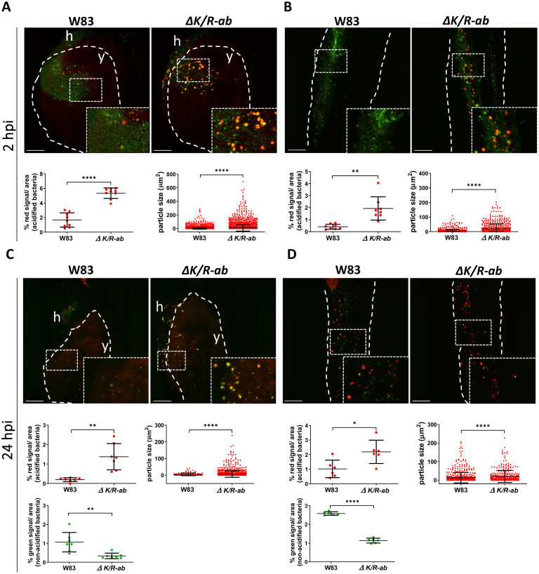
Gingipains prevent phagosome acidification of internalized Porphyromonas gingivalis (Pg).

Real-time in vivo examination of the acidification of the wild-type Pg W83 and ΔK/R-ab. Pg were stained with fluorescein-SE (green) and pHrodo (red) and zebrafish larvae (30 hpf) were infected into the Duct of Cuvier. Representative images along with quantification of the phagosome-acidified bacteria within selected area and size of acidified bacterial clusters in the yolk (A and C) or the tail regions (B and D) at 2 hpi (A and B) or 24 hpi (C and D). White dashed lines indicate yolk or tail areas. Boxes show zoomed regions of the images. y = yolk, h = heart. Scale bar = 100 μm. Data presented are means ± SD. Each dot represents signal quantified from a single zebrafish larva. Statistical significance was analysed by unpaired t-test with Welch’s correction or Mann-Whitney test where required. *≤0.05, **p≤0.01, ****p≤0.0001.

Acknowledgments
This image is the copyrighted work of the attributed author or publisher, and ZFIN has permission only to display this image to its users. Additional permissions should be obtained from the applicable author or publisher of the image. Full text @ PLoS Pathog.