
Fig. 1
- ID
- ZDB-IMAGE-250204-53
- Publication
- Wang et al., 2024 - Integrated transcriptomic analysis reveals evolutionary and developmental characteristics of tendon ossification in teleost
- All Figures
- Figures for Wang et al., 2024
|
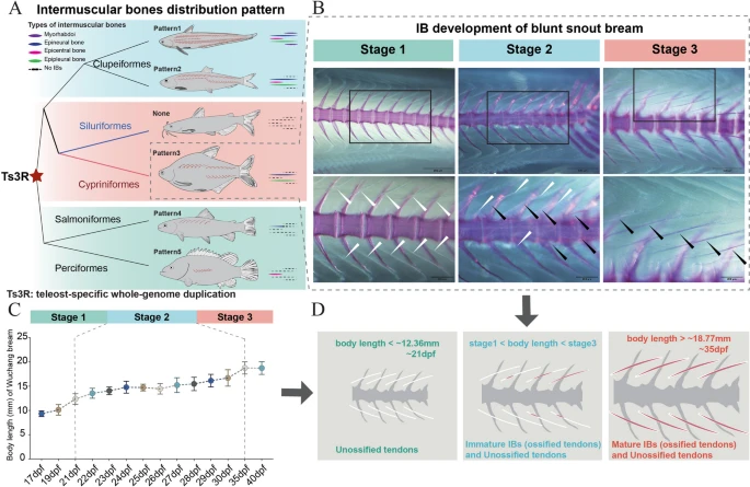
Fig. 1 The developmental and evolutionary characteristics of IBs (tendon ossification in myosepta). A IB distribution patterns in teleosts. Pattern 1 (Clupeiformes, Japanese grenadier anchovy (Coilia nasus)), epineural bones (ENBs), epipleural bones (EPBs), epicentral bones (ECBs), and myorhabdois (MBs); pattern 2 (Clupeiformes, Atlantic herring (Clupea harengus)), ENBs, EPBs, ECBs; pattern 3 (Cypriniformes, blunt snout bream (M. amblycephala)): ENBs and EPBs; none (Siluriformes, channel catfish (Ictalurus punctatus)), no IBs; pattern 4 (Salmoniformes, rainbow trout (Oncorhynchus mykiss)): ENBs; pattern 5 (Perciformes, Mandarin fish (Siniperca chuatsi)): ECBs. B–D Three developmental stages of IBs in blunt snout bream. B Histological analysis of IB development. White arrows marker tendons in myosepta; black arrows marker ossified tendons (IBs) in myosepta. C The relationship between IB development and body length. D Stage 1: only unossified tendon distribution without IB development (body length < ~ 12.36 mm); stage 2: distribution of both immature IBs (ossified tendons) and unossified tendons (body length ~ 12.36– ~ 18.77 mm); stage 3: distribution of mature IBs (ossified tendons) (body length > 18.77 mm)