IMAGE

Fig. 3

ID
ZDB-IMAGE-250203-20
Source
Figures for Meng et al., 2025
Image
Figure Caption

Fig. 3

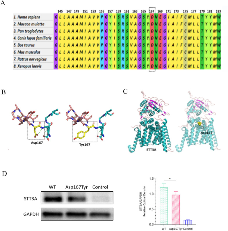
Characteristics of the variant and protein levels of STT3A. A Conservation analysis of this variant, which is conserved across multiple species. B Molecular modeling of the variant indicated a reduction in hydrogen bonds around the residue. C Molecular modeling of STT3A showing that the heterozygous missense variant is located within the binding site. D Western blot analysis of whole-cell protein extracts from transfected HEK293T cells. Compared with that in the WT group, the STT3A expression level in the Asp167Tyr group was significantly lower

Acknowledgments
This image is the copyrighted work of the attributed author or publisher, and ZFIN has permission only to display this image to its users. Additional permissions should be obtained from the applicable author or publisher of the image. Full text @ Orphanet J Rare Dis