IMAGE

Fig. 2

ID
ZDB-IMAGE-250203-19
Source
Figures for Meng et al., 2025
Image
Figure Caption

Fig. 2

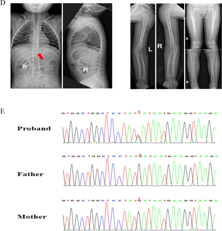
Auxiliary examination of the proband. A The peripheral blood flow cytometry results of the proband indicated a significant decrease in CD107a in the NK cells. B Video electroencephalogram revealed that the proband had abnormal discharges. In the awake state, moderate theta activity was detected at 4–7 Hz in the background without an occipital dominant rhythm. During wakefulness and sleep, spike-slow wave or slow wave bursts were observed in the left parietal and posterior temporal regions, which spread diffusely. C Brain magnetic resonance imaging (MRI) revealed an arachnoid cyst in the occipital region, along with bilateral enlarged lateral ventricles. D Spinal X-rays showing mild scoliosis with a Cobb angle of 4°, as indicated by the arrows. Limb X-rays revealed no abnormalities. E The Sanger sequencing of trios

Acknowledgments
This image is the copyrighted work of the attributed author or publisher, and ZFIN has permission only to display this image to its users. Additional permissions should be obtained from the applicable author or publisher of the image. Full text @ Orphanet J Rare Dis