
|
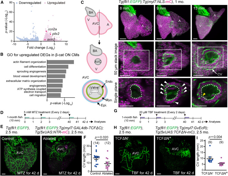
Fig. 3 β-cat ON CMs contribute to CV formation (A) A volcano plot showing differentially expressed genes (DEGs) between β-cat ON and OFF CMs (n = 3). Note that Wnt target genes (red circle) were upregulated. (B) Gene Ontology (GO) enrichment analysis of genes upregulated in β-cat ON CMs. (C) Schematic illustration of the heart (left) and representative images of the hearts of 1-month-old Tg(fli1:EGFP);Tg(myl7:NLS-mCherry) fish with body length indicated at the top (right). Hearts were made transparent with CUBIC-1A. Lateral projections (top), 50 μm stacked images of the dorsal view around the AVC (middle), and single slice images of the boxed regions (bottom). Dotted lines outline the heart lumen. Endothelial cells (ECs) are observed in the CM layer (white arrowheads). Coronary vessels (CVs) connect with endoECs at the AVC (yellow arrowhead). (D) Experimental design for specific ablation of nitroreductase-expressing (NTR-expressing) cells of the fish used in (E) by metronidazole (MTZ) treatment. 1-month-old fish were treated with 5 mM MTZ every 2 days and analyzed after 42 days of treatment. (E) Representative images of the CVs of Tg(fli1:EGFP) or Tg(fli1:EGFP);Tg(myl7:GAL4db-TCFΔC);Tg(5xUAS:NTR-mCherry) fish at 2.5 months treated with MTZ. A control heart (left) and a β-cat ON CM-ablated heart (right). (F) Quantitative analysis of the data shown in (E). The length of CVs was measured by tracing the length of fli1:EGFP+ coECs. (G) Experimental design for CM-specific induction of dominant negative TCF (TCFΔN) by tebufenozide (TBF) treatment in the fish used in (H). Fish were treated with 20 μM TBF every 2 days and analyzed after 42 days. (H) Representative images of the CVs of Tg(fli1:EGFP) or Tg(fli1:EGFP);Tg(myl7:GvEcR);Tg(5xUAS:TCFΔN-mCherry) fish at 2.5 months treated with TBF. A control heart (left) or a TCFΔN-expressed heart (right). (I) Quantitative analysis of the data shown in (H). Scale bars, 100 μm. Data are mean ± SEM. Statistical analysis was performed by two-tailed Student’s t test (F) or Mann-Whitney U test (I). CV, coronary vessel; Endo, endocardium; Epi, epicardium; Myo, myocardium. See also Figure S3, Table S1, and Video S1C.
Reprinted from Developmental Cell, 60(1), Chiba, A., Yamamoto, T., Fukui, H., Fukumoto, M., Shirai, M., Nakajima, H., Mochizuki, N., Zonated Wnt/β-catenin signal-activated cardiomyocytes at the atrioventricular canal promote coronary vessel formation in zebrafish, 21-29.e8, Copyright (2024) with permission from Elsevier. Full text @ Dev. Cell