
|
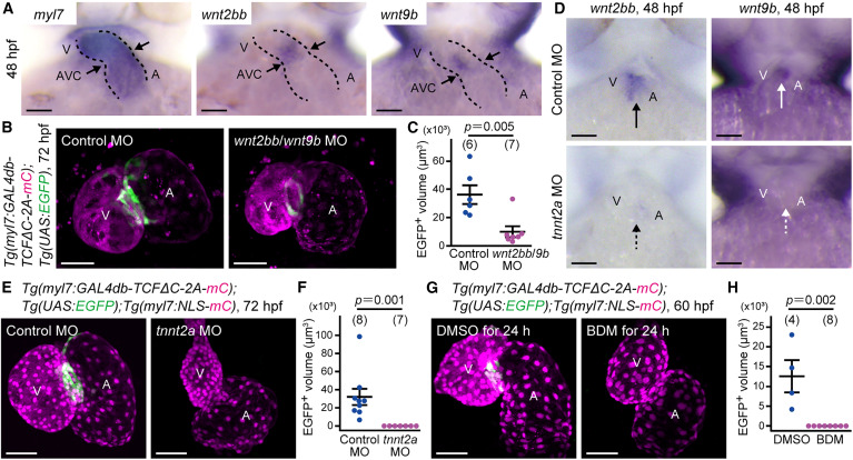
Fig. 2 Heartbeat-dependent Wnt expression turns on Wnt/β-catenin signaling in AVC CMs (A) Representative stereomicrographs of whole-mount in situ hybridization (WISH) analyses of myl7 (left), wnt2bb (center), and wnt9b (right) mRNA expression in three zebrafish embryos at 48 hpf. Ventral views. Dotted lines outline the heart. Arrows indicate the AVC. (B) Representative images of the hearts of Tg(myl7:GAL4db-TCFΔC-2A-mCherry);Tg(UAS:EGFP) larvae at 72 hpf injected with 7 ng control morpholino oligonucleotide (MO) (left) and 5 ng wnt2bb and 2 ng wnt9b MO (right). (C) Quantitative analysis of the data shown in (B). The volume of the UAS:EGFP+ region was measured. In this and the following graphs, unless otherwise described, each dot represents an individual fish, and the number of fish analyzed is indicated at the top. (D) Representative stereomicrographs of WISH analyses of wnt2bb (left) and wnt9b (right) mRNA expression in four embryos at 48 hpf. Embryos were injected with 2 ng control MO (top) and 2 ng tnnt2a MO (bottom). Arrows and broken arrows indicate positive and less mRNA expression, respectively. (E) Representative images of the hearts of Tg(myl7:GAL4db-TCFΔC-2A-mCherry);Tg(UAS:EGFP);Tg(myl7:NLS-mCherry) larvae at 72 hpf injected with 2 ng control MO (left) and 2 ng tnnt2a MO (right). (F) Quantitative analysis of the data shown in (E). (G) Representative images of the hearts of Tg(myl7:GAL4db-TCFΔC-2A-mCherry);Tg(UAS:EGFP);Tg(myl7:NLS-mCherry) embryos at 60 hpf treated with DMSO (left) or 20 mM 2,3-butanedione 2-monoxime (BDM) (right) for 24 h. (H) Quantitative analysis of the data shown in (G). Scale bars, 50 μm. Data are mean ± SEM. Statistical analysis was performed by Mann-Whitney U test. See also Figure S2.
Reprinted from Developmental Cell, 60(1), Chiba, A., Yamamoto, T., Fukui, H., Fukumoto, M., Shirai, M., Nakajima, H., Mochizuki, N., Zonated Wnt/β-catenin signal-activated cardiomyocytes at the atrioventricular canal promote coronary vessel formation in zebrafish, 21-29.e8, Copyright (2024) with permission from Elsevier. Full text @ Dev. Cell