
|
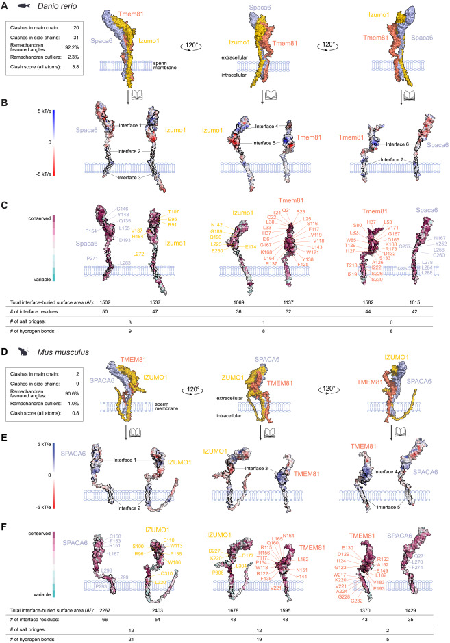
Fig. s2 Structural analysis of the interfaces of the predicted zebrafish and murine trimeric complexes, related to Figure 1 (A and D) Surface representation of the AlphaFold2-Multimer (v2)-predicted zebrafish (A) and murine (D) IZUMO1-TMEM81-SPACA6 complex shown in three orientations highlighting each binary interaction. Key structural properties of the model are quantified (left box). (B) Seven contact interfaces are found at the Izumo1-Spaca6-Tmem81 complex. Spaca6 and Izumo1 interact via two interfaces in the extracellular domain and one interface in the transmembrane. In Izumo1-Tmem81, two interfaces in the extracellular domain form contacts, while Tmem81-Spaca6 forms interactions between its extracellular and transmembrane domains. Electrostatic potential surface representation of the complex shows strong electrostatic complementarity at the interfaces. Residues colored in red and blue indicate electronegative and electropositive regions, respectively. Electrostatic potentials (in units of kT/e) were calculated using the program APBS.61 (C and F) Surface residue conservation of zebrafish (C) and murine (F) trimers. The degree of residue conservation is calculated in comparison with mammalian orthologs using the program Scorecons62 and shown in a gradient from high (cyan) to low (magenta) variability. Residues that are strictly conserved in mammals are listed in the figure. The footprints of the binding interfaces are shown by solid black lines. Tables provide the total interface-buried surface area and the number of interface residues, salt bridges, and hydrogen bonds per interacting protein pair. (E) Five contact interfaces are found in the murine IZUMO1-SPACA6-TMEM81 complex. SPACA6-IZUMO1 and TMEM81-SPACA6 interact via interfaces in each of their extracellular and transmembrane domains, whereas in IZUMO1-TMEM81, the extracellular domains form a single interface. Electrostatic potential surface representation of the murine IZUMO1-SPACA6-TMEM81 complex show strong electrostatic complementarity at the interfaces. See (B) for further details. All figures were generated in PyMOL (Schrodinger).
Reprinted from Cell, 187(25), Deneke, V.E., Blaha, A., Lu, Y., Suwita, J.P., Draper, J.M., Phan, C.S., Panser, K., Schleiffer, A., Jacob, L., Humer, T., Stejskal, K., Krssakova, G., Roitinger, E., Handler, D., Kamoshita, M., Vance, T.D.R., Wang, X., Surm, J.M., Moran, Y., Lee, J.E., Ikawa, M., Pauli, A., A conserved fertilization complex bridges sperm and egg in vertebrates, 7066-7078.e22, Copyright (2024) with permission from Elsevier. Full text @ Cell