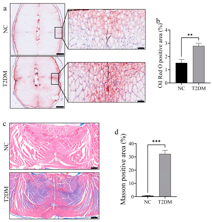
IMAGE

Figure 3

ID
ZDB-IMAGE-250127-145
Source
Figures for Chen et al., 2025
Image
Figure Caption

Figure 3

Lipid accumulation and fibrosis were significantly enhanced in the skeletal muscle of zebrafish with type 2 diabetes mellitus (T2DM). (a) Oil red O staining of skeletal muscle after 8 weeks of high-glucose treatment, with red indicating fat-positive staining. The image on the left is a low-magnification image (scale bar: 500 μm); the picture on the right is an enlarged view of the slow-twitch muscle area (scale bar: 50 μm). (b) Positive area of Oil red O staining; (c) Masson staining intensity in the skeletal muscle (scale bar: 200 μm); and (d) statistical chart of Masson staining positive area. ** p < 0.01, *** p < 0.001. (n = 3 samples/group).

Acknowledgments
This image is the copyrighted work of the attributed author or publisher, and ZFIN has permission only to display this image to its users. Additional permissions should be obtained from the applicable author or publisher of the image. Full text @ Int. J. Mol. Sci.