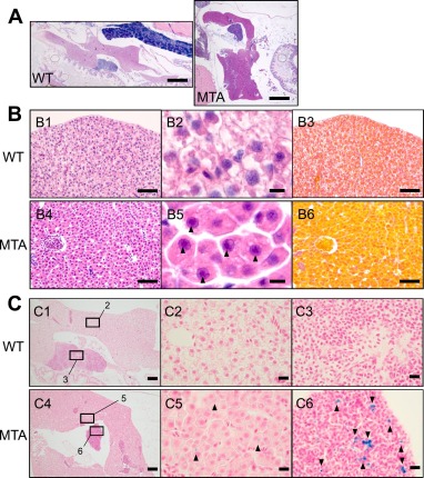
Fig. 6 Abnormal appearance of hepatocytes and iron depositions in the liver and spleen of the slc25a20 mutant. (A) Representative images of the whole liver from hematoxylin and eosin (HE)-stained paraffin sections of the individuals. Scale bar, 0.5 mm. (B) Representative images of liver tissues from the wild-type fish (B1–3) and the mutant (B4–6). (B1, B4) Low-magnification images of the livers from HE-stained paraffin sections. Scale bars, 50 μm. (B2, B5) High-magnification images of hepatocytes. The back arrowheads indicate prominent nucleoli. Scale bars, 5 μm. (B3, B6) Images of azan-stained paraffin sections adjacent to those in B1 and B4. (C) Representative images of the liver and spleen from Prussian blue-stained paraffin sections of the wild-type fish (C1–3) and the mutant (C4–6). (C1, C4) Low-magnification images. Squares correspond to the magnified regions shown in the following images. Scale bars, 100 μm. (C2, C5) High-magnification images of the livers. Scale bars, 10 μm. (C3, C6) High-magnification images of the spleens. The back arrowheads indicate focal trace iron depositions. Scale bars, 10 μm.
Image
Figure Caption
Figure Data
Acknowledgments
This image is the copyrighted work of the attributed author or publisher, and
ZFIN has permission only to display this image to its users.
Additional permissions should be obtained from the applicable author or publisher of the image.
Full text @ Mol Genet Metab Rep

