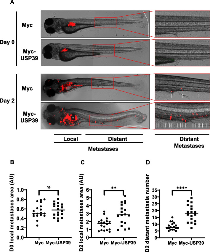
USP39 Enhances Metastasis in Zebrafish: Implications for MM Progression. A-D Zebrafish embryos (N = 36) were injected with U266 cells stably infected with lentiviral particles encoding either Myc-tag or Myc-USP39 proteins (labeled with red DiD) into the perivitelline space. Zebrafish embryos were monitored on Day 0 and Day 2 for tumor metastases using a fluorescent microscope. A (Left) Representative images of local and distant metastases are shown. (Right) Magnification of images representing distant metastases. Arrows indicate metastases. Quantification of the area of local metastases at Day 0 (B) and Day 2 (C) of Myc-tag and Myc-USP39 embryos. D Quantification of the number of distant metastases at Day 2 of Myc-tag and Myc-USP39 embryos
Acknowledgments
This image is the copyrighted work of the attributed author or publisher, and
ZFIN has permission only to display this image to its users.
Additional permissions should be obtained from the applicable author or publisher of the image.
Full text @ J. Exp. Clin. Cancer Res.
Your Input Welcome
Thank you for submitting comments. Your input has been emailed to ZFIN curators who may contact you if
additional information is required.
Oops. Something went wrong. Please try again later.