
|
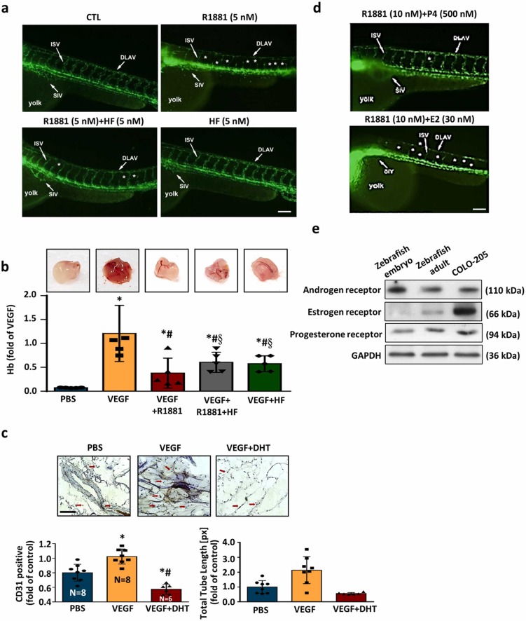
Fig. 7 R1881 attenuates angiogenesis and modulates the impact of female sex hormones on R1881-induced anti-angiogenic responses. (a) Zebrafish angiogenesis model was conducted to evaluate the effect of R1881 on angiogenesis. Treatment with R1881 for 72 h after post-fertilization caused failure in forming the ISV and the DLAV in embryos. However, co-treatment with HF abolished the R1881-induced interruption of angiogenesis. Fluorescent images show defective vasculatures in ISV and DLAV at the R1881-treated Tg(fli1:EGFP)y1 embryos. The white arrowheads indicate the failure of forming the ISV or DLAV in the R1881-treated Tg(fli1:EGFP)y1 embryos. Bar = 100 μm. (b) The effect of R1881 on angiogenesis was evaluated using matrigel plug assay as described in the Materials and Methods section. Treatment with VEGF (200 ng/mL) for 14 days increased the level of Hb, an indicator of angiogenesis. Top panel shows the matrigel isolated from the C57BL/6 mouse. Bottom panel shows the quantitative results of Hb. (c) DHT (5 nM) reduced the VEGF-increased angiogenesis. The matrigel plug isolated from the mouse was cut into 5 μm sections and stained with an anti-CD31 antibody. The arrowheads indicate the positive CD31 immunoreactivity (endothelial cell marker). Bar = 100 μm. Top panel shows a representative section stained with an anti-CD31 antibody. Bottom panel shows the quantitative results of CD31 positive cell number. (n = 5). Values present the means of fold of corresponding control ± s.e.mean. (n = 4). *p < 0.05 different from PBS group (control). #p < 0.05 different from the VEGF-treated group. §p < 0.05 different from the VEGF+R1881-treated group. DLAV, dorsal longitudinal anastomotic vessels; Hb, hemoglobin; ISV, intersegmental vessels. (d) Treatment with R1881 at 20 h after post-fertilization significantly inhibited angiogenesis and this inhibition was suppressed by co-treatment with HF (5 nM). Co-treatment with P4 (500 nM), but not E2 (30 nM), prevented the R1881-induced anti-angiogenesis. Embryos were examined at 2 days post-fertilization. More than 20 embryos were examined in each experimental group. Twenty dechorionated Tg(flil:EGFP)y1 embryos were grown in a 24-well plate, and incubated in 2 mL solution containing R1881 (10 nM). Fluorescent images show defective vasculatures in ISV (arrowheads) and DLAV (arrows) at the R1881-treated Tg(flil:EGFP)y1 embryos. The stars indicate the failure of forming the ISV and DLAV in the R1881-treated Tg(flil:EGFP)y1 embryos. Bar = 100 μm. (e) The expressions of AR, PR and ER were detected in the adult zebrafish at age of older than 1 year, but only AR and PR were detected in zebrafish embryos at 20 h post-fertilization. AR, androgen receptor; CTL, control; DLAV, dorsal longitudinal anastomotic vessels; ER, estrogen receptor; ISV, intersegmental vessels; NC, notochord; PR, progesterone receptor.