
|
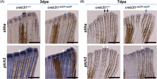
Fig. 8 creb3l1 mutant fish exhibit aberrant patterning of shha and ptch2. (A, B) Approximately 50% of the caudal fin was amputated from 6 mpf creb3l1+/+ and creb3l1ΔbZIP/ΔbZIP fish. Regenerates at 3 dpa (A) and 7 dpa (B) were analyzed by in situ hybridization to assess the expression patterns of shha and ptch2. Amputation planes are indicated with dashed white line. Scale bar = 1 mm. (A) The shha signal in wild-type creb3l1+/+ fish exhibits the characteristic two-point staining (black arrows). In contrast, in creb3l1 mutant fish, shha signal is largely in a single domain over the regenerating ray (blue arrow). The ptch2 signal in wild-type creb3l1+/+ regenerate is segregated into defined caps over regenerating rays with unstained areas between the caps (white arrow). In contrast, patch2 expression is more diffuse in creb3l1ΔbZIP/ΔbZIP regenerate (white arrow). (B) In wild-type creb3l1 fish, the shha signal appears in two clearly separated foci over each bifurcated ray (black arrows). In contrast, shha signal is more diffuse in creb3l1 mutant regenerates (blue arrow). The ptch2 signal in wild-type creb3l1+/+ fish segregates into two domains over each bifurcated ray (white arrows). In contrast, in creb3l1ΔbZIP/ΔbZIP mutant regenerate, ptch2 signal predominantly remains within a single diffuse domain (white arrows). n = 3–7.