
Image
|
Figure Caption
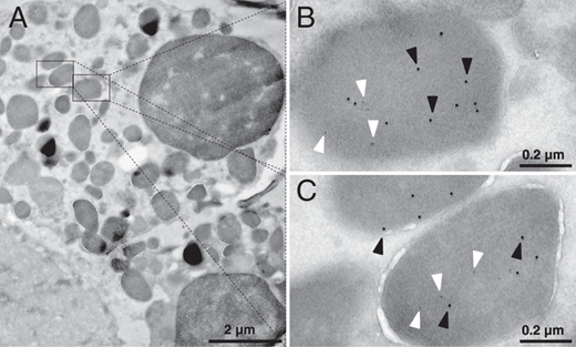
Fig. 5 Cryo-immunogold EM of FACS isolated Embp-tdTomato+ eosinophils. (A) Overview of an eosinophil. (B and C) Zoom in on two different granula of this cell. Embp Ab is visualized by a 12-nm gold secondary Ab (black arrowheads) and mCherry Ab is visualized by 6-nm gold secondary Ab (white arrowheads). Both target Ags were detected in the same granule.
Acknowledgments
This image is the copyrighted work of the attributed author or publisher, and
ZFIN has permission only to display this image to its users.
Additional permissions should be obtained from the applicable author or publisher of the image.
Full text @ J. Immunol.