
|
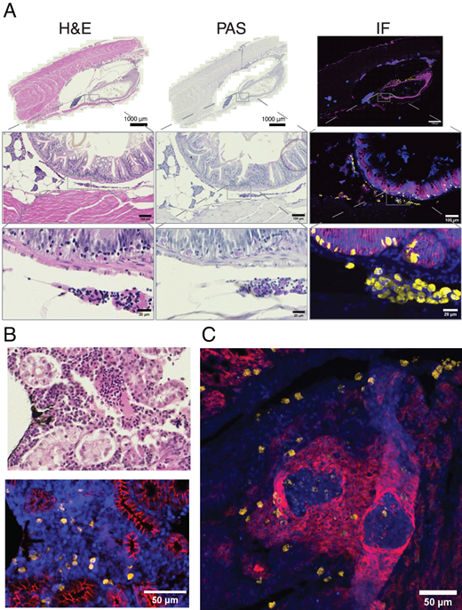
Fig. 4 Embp Ab staining in FFPE tissue samples. (A) H&E, PAS, and immunofluorescence (IF) staining with Embp Ab in healthy TgKI(embp-tdTomato,cryaa:EGFP) adults. Blue indicates DAPI; yellow indicates eosinophils; magenta indicates E-cadherin staining of epithelial cells. n = 3. (B) Zoom in on a part of the WKM, showing the Embp+ stain in this tissue together with the corresponding site in H&E staining of the adjacent section. Same IF staining as in (A). (C) Staining of eosinophils in M. marinum–infected zebrafish FFPE sections. Eosinophils can be found in physiological sites in the body as well as in granuloma. Magenta indicates E-cadherin, marking epithelioid macrophages surrounding the necrotic core of the granuloma; yellow indicates eosinophils stained by Embp Ab; blue indicates DAPI.