IMAGE

Figure 2

ID
ZDB-IMAGE-241219-101
Source
Figures for Hsu et al., 2024
Image
Figure Caption

Figure 2

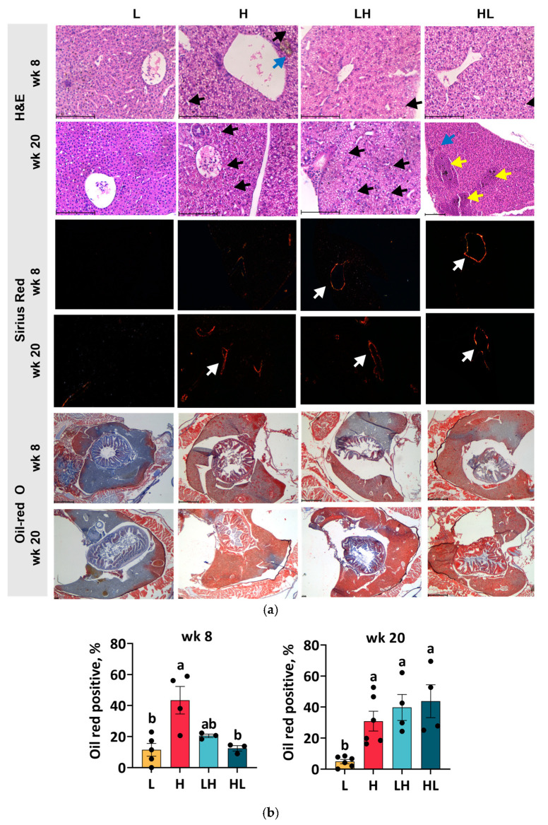
Liver histology. The liver sections were individually stained with H&E, Sirius red, and Oil Red O (a). The positive area of oil red-O staining at week 8 and week 20 was quantified (b). The scale bar for H&E and Sirius red is 100 µm, and that for Oil Red O is 500 µm. Black arrows indicate the ballooning hepatocytes; blue arrows indicate the cholestasis site; yellow arrows indicate hyperemia; and white arrows indicate fibrosis. ab In the graphs, lines, and bars sharing different letters are significantly different (p ≤ 0.05). Data are presented as the mean ± SEM (n = 3–6).

Acknowledgments
This image is the copyrighted work of the attributed author or publisher, and ZFIN has permission only to display this image to its users. Additional permissions should be obtained from the applicable author or publisher of the image. Full text @ Int. J. Mol. Sci.