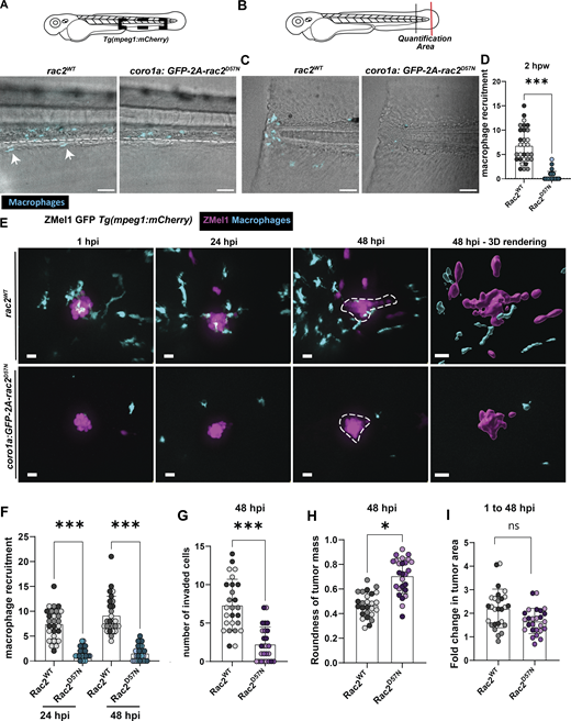
Fig. 3 Rac2 signaling in macrophages is required for macrophage recruitment to the TME and tumor invasion. (A) Representative images of macrophages in the caudal hematopoietic tissue of 3 dpf wild-type larvae or Tg(coro1a:GFP-rac2 D57N)psi92T larvae. (B) Schematic of a 3 dpf larvae to highlight the tail transection region (red line) and the quantification area for macrophage recruitment (between red and black line). (C) Representative images of the tail fin of wild-type or Rac2D57N larvae wounded and imaged 2 h post wound. (D) Quantification of macrophage recruitment to tail fin. n = 30 wild-type, n = 27 Rac2D57N from three independent replicates. (E) Representative images from time course imaging of wild-type or Rac2D57N larvae injected with Zmel1 GFP cells from 1 hpi to 48 hpi. (F) Quantification of macrophage recruitment to tumor at 24 and 48 hpi. (G) Quantification of number of invaded tumor cells at 48 hpi. (H) Quantification of tumor roundness at 48 hpi. (I) Quantification of fold change in tumor area from 1 to 48 hpi. n = 26 wild-type, n = 26 Rac2D57N from three independent replicates. Scale bar = 20 µm. *P < 0.05, **P < 0.01, ***P < 0.001, n.s., not significant.
Image
Figure Caption
Acknowledgments
This image is the copyrighted work of the attributed author or publisher, and
ZFIN has permission only to display this image to its users.
Additional permissions should be obtained from the applicable author or publisher of the image.
Full text @ J. Cell Biol.

