IMAGE

Fig. 4

ID
ZDB-IMAGE-241209-9
Genes
Source
Figures for Park et al., 2024
Image
Figure Caption

Fig. 4

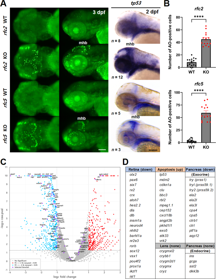
Increased cell death in rfc2 KO and rfc5 KO zebrafish. A: Detection of cell death by AO staining in KO zebrafish at 3 dpf. Apoptotic cells were mainly detected in midbrain, mhb, and retina. Earlier apoptotic cells were confirmed by the apoptosis marker tp53 expression at 2 dpf. Left panels are dorsal view, middle and right panels are lateral view, anterior is to the left. B: Quantification of AO-positive cells within the brain region in rfc2 KO or rfc5 KO zebrafish. n = 16 for rfc2 WT, n = 14 for rfc2 KO, n = 15 for rfc5 WT, and n = 14 for rfc5 KO. Data are presented as mean ± SEM. Statistical significance was determined by the unpaired two-tailed t-tests with Welch's correction. ∗∗∗∗, P < 0.0001. C: Visualization of RNA-seq analysis with volcano plot. Blue dots represent down-regulated, red dots for up-regulated, gray dots for not significantly changed, and purple dots for labeled genes in rfc2 KO, compared to WT. D: List of representative gene sets involved in eye development or pancreas function. Scale bar, 100 μm (A). AO, acridine orange; mhb, midbrain-hindbrain boundary; WT, wild-type; KO, knockout; SEM, standard error of the mean.

Figure Data
Acknowledgments
This image is the copyrighted work of the attributed author or publisher, and ZFIN has permission only to display this image to its users. Additional permissions should be obtained from the applicable author or publisher of the image.

Reprinted from Journal of genetics and genomics = Yi chuan xue bao, 51(12), Park, J.W., Choi, T.I., Kim, T.Y., Lee, Y.R., Don, D.W., George-Abraham, J.K., Robak, L.A., Trandafir, C.C., Liu, P., Rosenfeld, J.A., Kim, T.H., Petit, F., Kim, Y.M., Cheon, C.K., Lee, Y., Kim, C.H., RFC2 may contribute to the pathogenicity of Williams syndrome revealed in a zebrafish model, 1389-1403, Copyright (2024) with permission from Elsevier. Full text @ J. Genet. Genomics