IMAGE

Fig. 2

ID
ZDB-IMAGE-241209-17
Source
Figures for Begum et al., 2024
Image
Figure Caption

Fig. 2

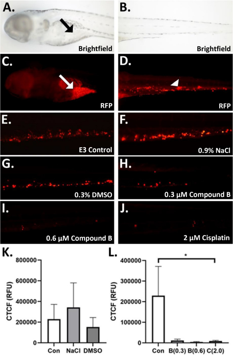
Compound B reduces ovarian cancer metastasis comparably to cisplatin.Images show ovarian cancer cells labeled with DiI membrane stain visible in the red fluorescent channel. A.-D. Representative images of microinjected zebrafish embryos (A. brightfield cranial view showing yolk sac injection site (black arrow); B. brightfield caudal view; C. red channel fluorescent cranial view showing yolk sac injection site with cancer cells (white arrow); D. red channel fluorescent caudal view showing metastasized A2780cis cells (white arrow head), E.-J. Representative images of control and experimental treatment zebrafish (E., E3 media control; F., 0.9% NaCl cisplatin control; G., 0.3% DMSO compound B control; H., 0.3 µM compound B; I., 0.6 µM compound B; J., 2.0 µM cisplatin). K. Comparison of E3 media (Con), 0.9% NaCl, and 0.3% DMSO controls. L. Comparison of E3 control (Con) with three platinum complex treatments: 0.3 µM compound B [B(0.3)], 0.6 µM compound B [B(0.6)], 2.0 µM cisplatin [C(2.0)]. CTCF = corrected total cell fluorescence; p < 0.05, “*” = 0.05; N = 5–15

Acknowledgments
This image is the copyrighted work of the attributed author or publisher, and ZFIN has permission only to display this image to its users. Additional permissions should be obtained from the applicable author or publisher of the image. Full text @ Invest New Drugs