IMAGE

Fig. 1

ID
ZDB-IMAGE-241205-1
Source
Figures for Bowley et al., 2024
Image
Figure Caption

Fig. 1

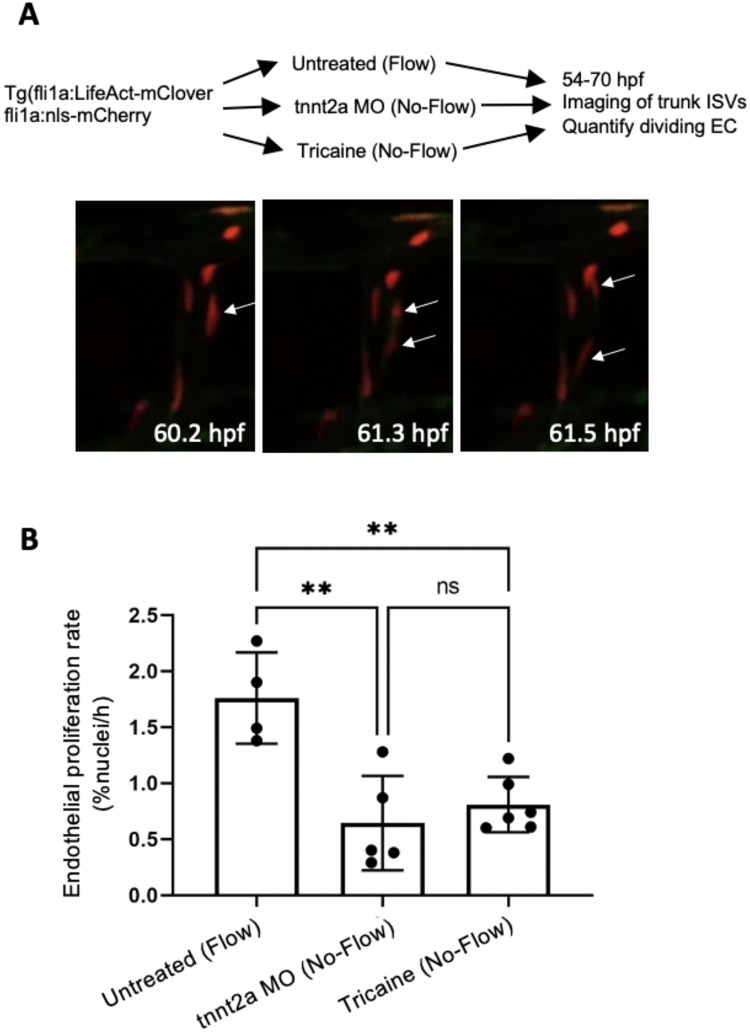
EC proliferation is significantly reduced in embryos without blood flow. Transgenic Tg(fli1a:LifeAct-mClover; fli1a:nls-mCherry) embryos were analysed. The ISVs of four embryos with flow, five tnnt2a-MO (No-Flow), and six tricaine-treated (No-Flow) embryos were imaged from 54 to 70 hpf. For each embryo, data were pooled from 7 ISVs. (A) Representative images showing a dividing EC (arrows) in an ISV exposed to flow. (B) The rate of EC proliferation in the ISVs was quantified as a percentage of nuclei dividing per hour. Mean values + /− standard deviations are shown. Differences between means were analysed using an one-way ANOVA with multiple comparisons.

Acknowledgments
This image is the copyrighted work of the attributed author or publisher, and ZFIN has permission only to display this image to its users. Additional permissions should be obtained from the applicable author or publisher of the image. Full text @ Sci. Rep.