
|
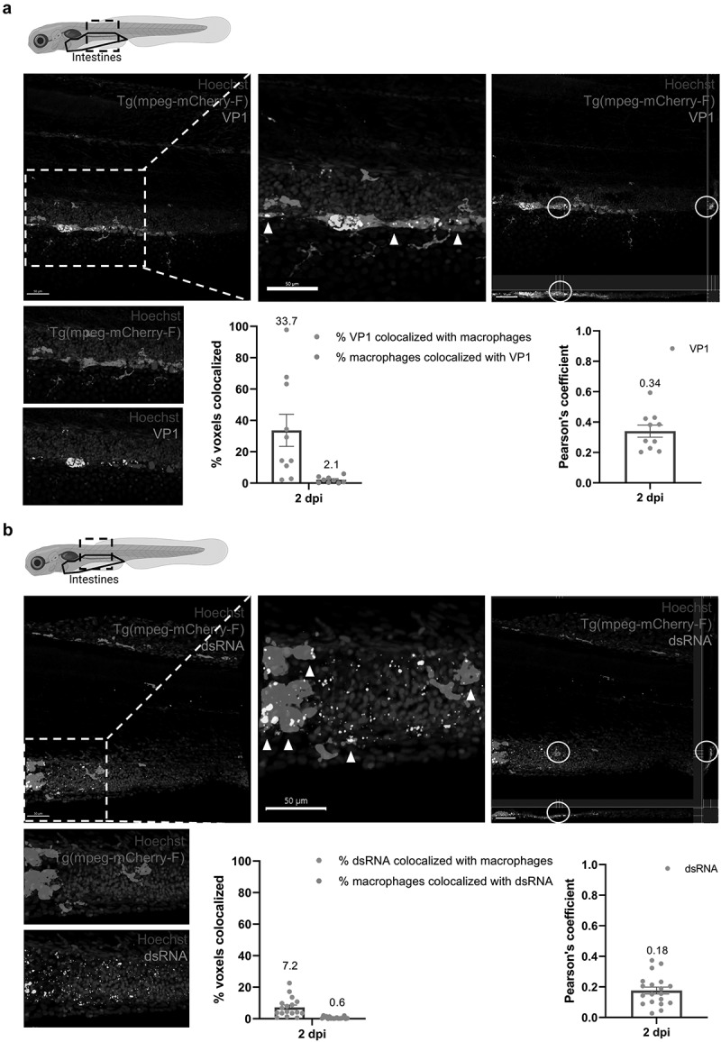
Figure 4.
Macrophages in the intestines are infected by HuNoV and allow active viral replication. (a-b) whole mount immunohistochemistry confocal images of Tg(mpeg:mCherry-F) zebrafish larvae infected with HuNoV taken at 2 dpi at a 25X magnification focusing on the intestines using Hoechst, a primary mCherry antibody, and (a) a VP1-targeting antibody or (b) a dsRNA-targeting antibody. Cross-section views created in imaris software show horizontal and vertical sections of macrophages that contain (a) VP1 or (b) dsRNA with the white circle highlighting an infected cell of interest. Imaris 3D Colocalization software calculated the overlap of voxels between macrophages and (a) VP1 or (b) dsRNA (