IMAGE

Figure 4.

ID
ZDB-IMAGE-241119-98
Source
Figures for Tanaka et al., 2024
Image
Figure Caption

Figure 4.

Depletion of GnRH3 neurons leads to reduced gonadotropes number in mature female pituitaries. WT females and Tg(gnrh3:Gal4ff; UAS:nfsb-mCherry) treated with either vehicle or Mtz for 14 days. Pituitaries were subjected to double labeling in situ hybridization followed by cell counting. (A) Pituitary of representative WT with Mtz (upper panel), Tg with vehicle (second panel from top), and Tg with Mtz (third panel from top) treated females. Fourth panel depicts a single 20 µm Z-stack image of WT pituitary stained with Hoechst 3342. From left to right: lhb mRNA (red), fshb mRNA (green), merged image to analyze colocalization of lhb and fshb, and a magnification of the rectangular area depicting region. (B) Percentage of lhb+, fshb+, and lhb+/fshb+ colocalized cells in the proximal pars distalis (PPD) counted in the photographed pituitaries (WT-Mtz and Tg-Cont, n = 5, Tg-Mtz, n = 4). Results are presented as mean ± SEM. Statistical significances were tested by one-way ANOVA followed by Tukey multiple comparison test. *P < .05, **P < .01, ***P < .001, ****P < .0001.

Abbreviations: Mtz, metronidazole; Cont, control; WT, wild-type; Tg, transgenic.
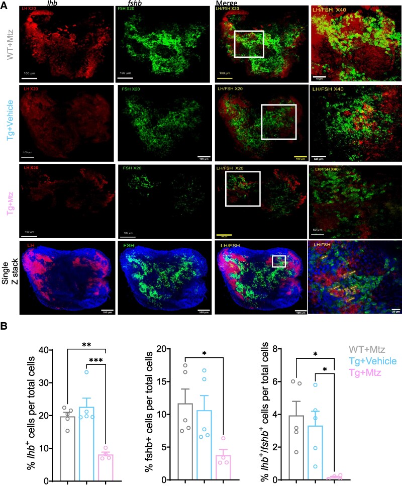
Acknowledgments
This image is the copyrighted work of the attributed author or publisher, and ZFIN has permission only to display this image to its users. Additional permissions should be obtained from the applicable author or publisher of the image. Full text @ Endocrinology