IMAGE

Fig 1

ID
ZDB-IMAGE-241114-28
Source
Figures for Yang et al., 2024
Image
Figure Caption

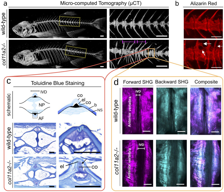
Fig 1 Effects of col11a2-/- knockout in zebrafish.

The col11a2 -/- zebrafish have a fused vertebral column, ruptured annulus fibrosus and morphological abnormalities. (a) Micro-computed Tomography (μCT) of 6-month wild–type and mutant fish. The region highlighted with dashed yellow box is shown with higher magnification on the right panel. Note vertebrae fusions (magenta dash lines). (b) Fluorescent microscopic images of the calcified tissues of zebrafish labelled by Alizarin Red staining. Note abnormal vertebral trabeculations (arrow) and bone outgrowths from the arches (dashed arrow). Scale bars = 500 μm. (c) Toluidine blue stained images showing the cellular components of the soft tissue intervertebral disc (IVD) region. The top schematic (left) shows the encounter of two vertebrae at the IVD region, with the annulus fibrosus (AF) and nucleus pulposus (NP) annotated. The top right schematic shows a higher magnification view of the annulus fibrosus (AF) region, which is formed by an external layer of collagen fibres (co), an elastin layer (el) and another collagen rich region (co). The vertebral bone (b) and the notochord sheath (ns) are annotated. The sections of the wild–type and col11a2 -/- fish are shown in the middle and bottom rows respectively. Note that the elastin (el) and the collagen (co) layers are ruptured (arrow). (d) Forward and backwards Second Harmonic Generation (SHG) images of the wild–type and col11a2 -/- zebrafish intervertebral disc area. Note thicker collagen fibres at the endplates (dashed line) of mutant fish. Disorganised collagen fibres in the IVD (AF) is detected in mutants (arrowhead) Scale bars = 50μm.

Acknowledgments
This image is the copyrighted work of the attributed author or publisher, and ZFIN has permission only to display this image to its users. Additional permissions should be obtained from the applicable author or publisher of the image. Full text @ PLoS Comput. Biol.