
|
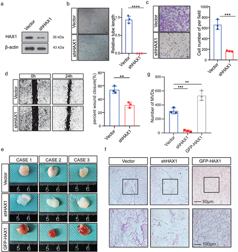
Fig. 4 HAX1 expression affects the tube formation ability of HUVECs. (a) HUVECs transfected with control (CON) or shHAX-1 were analysed by Western blotting. (b–d) HUVECs were transfected with the control (CON) or shHAX-1 lentivirus, followed by angiogenesis-related experiments. (b) Tube formation assays were performed to measure the tube forming ability of two groups of HUVECs, bar: 100µm. (****p < 0.0001, Student's t-test). (c) The migration potential of HUVECs was measured using the Transwell assay, bar: 100 µm. (**p < 0.01, Student's t-test) (d) The migration potential of HUVECs was measured using the wound closure assay, bar: 200 µm. (***p < 0.001, Student's t-test). (e, f) HUVECs were mixed with Matrigel for subcutaneous injection. (e) Left, gross-observation of HAX1-modulated angiogenesis in Matrigel plugs. Right, H&E was performed to observe blood vessel formation. Scale bar in 200× images: 100 µm. Scale bar in 400× images: 50 µm. (f) Quantitative evaluation of angiogenesis based on vessel area (**p < 0.01, ***p < 0.001, one-way ANOVA)