
|
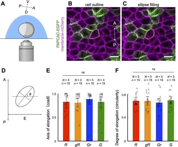
Fig. 2 PAPCΔC expressing cells polarize normally in the developing notochord. (A) A diagram showing the orientation of the embryo at the beginning of confocal microscopy (60% epiboly, see Methods). A–P and D–V indicate the anterior–posterior and dorsal–ventral axes, respectively. (B, C) Confocal images of axial mesodermal cells in an embryo expressing membrane-mCherry uniformly and PAPCΔC-EGFP in a mosaic fashion at 9 hpf. A–P indicates the anterior–posterior axis. Scale bars, 25 μm. (B) The outline of a cell (expressing membrane-mCherry and in contact with cells expressing PAPCΔC) is manually traced in yellow. (C) The outline of the same cell is fitted to an ellipse. (D) Evaluation of cell polarization via ellipse fitting. The axis of cell elongation was evaluated by the angle of the long axis of the ellipse (blue dotted line) to the equatorial line (E), which is perpendicular to the anterior–posterior (A–P) axis. The degree of cell elongation is evaluated by the circularity of the ellipse, which is determined by the length of the short axis (purple dotted line) divided by the long axis. (E, F) Axis of elongation (cosθ) (D) and degree of elongation (circularity) (E) of cells. Here, the cells were categorized into four categories according to their expression of PAPCΔC as well as contact with (an)other PAPCΔC-expressing cell(s): R, cells expressing mCherry only and surrounded by cells expressing mCherry only; gR, cells expressing mCherry only and in contact with PAPACΔC-expressing cell(s); Gr, cells expressing PAPCΔC and surrounded by cells expressing mCherry only; G, cells expressing PAPCΔC and in contact with (an)other PAPCΔC-expressing cell(s). Fifteen cells (n = 15) from three independent experiments (N = 3) were analyzed. ns, not significant (one-way ANOVA). The data are presented as the means ± s.d. Squares, circles and triangle represent single data points from three independent experiments.