
|
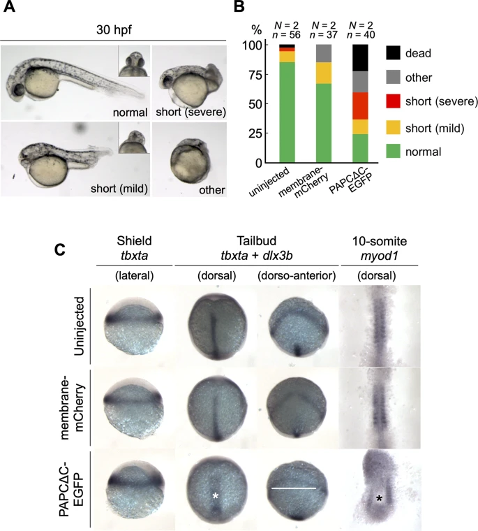
Fig. 1 The expression of PAPCΔC disrupts axis elongation during zebrafish development. (A) Embryos with various degrees of morphogenetic anomalies at 30 hpf. (Left top) A normal embryo. The inset shows the ventral view of the head region, with well-separated eyes. (Right top) A severe case of a shortened axis, classified by an axial length of approximately < 50% of that of a normal embryo, while both the anterior (eyes) and posterior (tail) structures are discernible. (Left bottom) A mild case of a shortened axis, classified by an axial length of approximately 50–80% of that of a normal embryo, accompanied by cyclopia (inset). (Right bottom) An embryo with severe developmental defects. (B) Morphogenetic phenotypes at 30 hpf. Fifty-six uninjected, 37 membrane-mCherry mRNA-injected, and 40 PAPCΔC-EGFP mRNA-injected embryos from two independent experiments were analyzed. (C) Whole mount in situ hybridization using probes for tbxta, dlx3b and myod1 at the shield, tailbud and 10-somite stages. The yolk was removed, and the embryos were mounted flat for 10-somite specimens. The asterisks and a white bar denote broader and shorter notochords and broader neural plate, respectively, in PAPCΔC-EGFP mRNA-injected embryos. Embryos from four independent experiments were analyzed.