
|
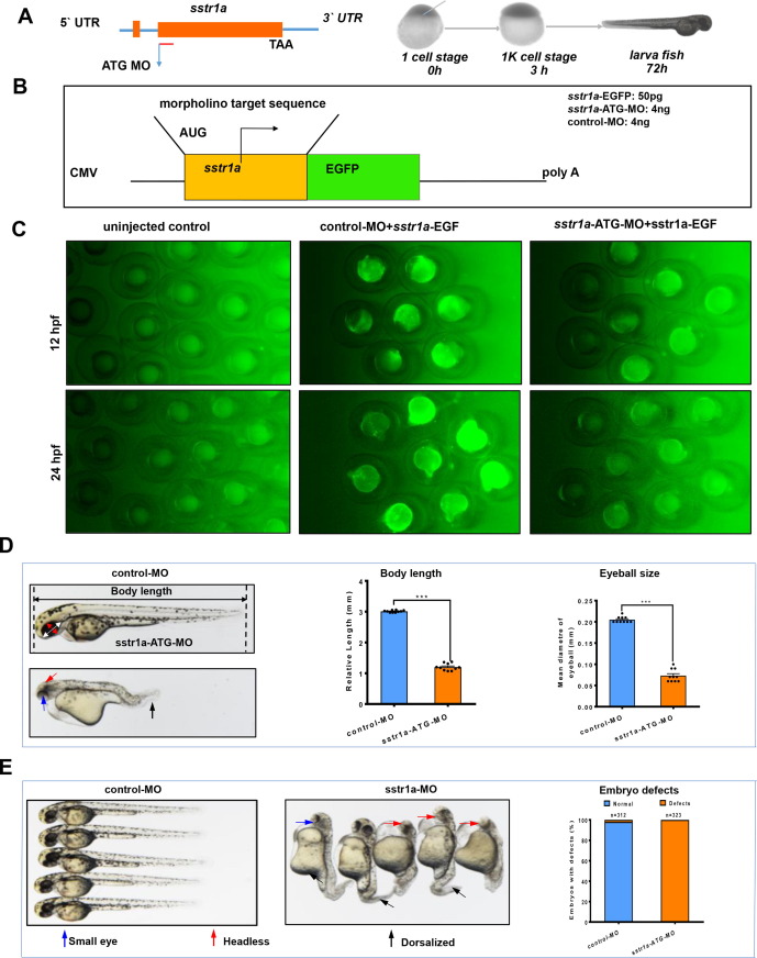
Fig. 7 Loss of sstr1a causes embryo defects in zebrafish. A. Schematic map of sstr1a gene and the MO design and micro-injection strategy. B. Site-specific effect of sstr1a-MO injected into zebrafish. Box shows schematic diagrams of sstr1a-EGFP fluorescent reporter mRNAs, the upper one containing the sstr1a-ATG-MO target sequence (yellow box) fused in-frame with EGFP. 50 pg sstr1a-EGFP injected with a standard control morpholino (4 ng) or sstr1a-ATG-MO (4 ng). C. Embryos were photographed at 12 and 24-hpf. Embryos injected with sstr1a-GFP plasmid DNA under the driving of CMV promoter showed green fluorescence. When co-injected with sstr1a-ATG-MO, green fluorescence decreased dramatically. hpf, hours post fertilization. D. Loss of sstr1a impacts the growth of zebrafish. Body length and eyeball size were measured at 2-dpf. E. Gross morphology at 2-dpf. Compared with control MO, knock down sstr1a present small eyes (blue arrow), tail patterning defects (dorsalized, black arrow), headless (red arrow) and pericardial oedema (purple arrow). The numbers of embryo defects were counted. Error bars, mean ± s.e.m.; ***p < 0.0001 (n = 10; Student’s t test). dpf, days post fertilization. (For interpretation of the references to color in this figure legend, the reader is referred to the web version of this article.)