
|
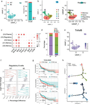
Fig. 7 Heterogeneity analysis of B cell subtypes. (a) UMAP analysis shows the distribution of B cells in different groups. Each point represents a single cell, colored by its subtype. (b) UMAP analysis shows the distribution of B cell subtypes in different samples. Each point represents a single cell. Different colors represent different B cell subtypes. (c) Bubble plots showing the expression of commonly used marker genes for each B cell subtype, used for annotating cell subtypes. The size of the bubble represents the percentage of cells expressing the marker gene, and the color intensity indicates the expression level.(d) Proportion of each B cell subtype in the WT and knockdown groups. (e) Expression of the B cell marker Tnfrsf8 (Cd30). (f) Differential genes in regulatory B cells between different groups. (g) Prognostic value of specific genes in regulatory B cells, based on survival analysis. A Two-tailed log-rank test was performed (h) Pseudotime analysis shows the developmental trajectory of B cell subtypes.