IMAGE

Figure 1

ID
ZDB-IMAGE-241019-28
Source
Figures for Wijerathna et al., 2024
Image
Figure Caption

Figure 1

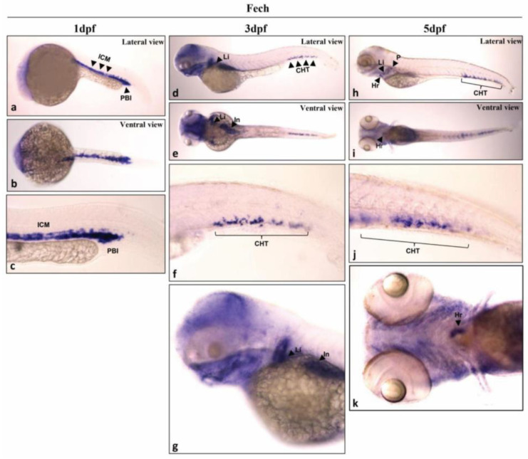
Spatial distribution of fech in zebrafish larvae. fech expression in larvae 1 day ((a) lateral view; (b) ventral view; and (c) tail), 3 days ((d) lateral view; (e) ventral view; (f) tail; (g) head), and 5 days ((h) lateral view; (i) ventral view; (j) tail; (k) head) days post-fertilization (dpf). ICM, intermediate cell mass; PBI, posterior blood island; Li, liver; CHT, caudal hematopoietic tissue; Hr, heart; P, pronephros; In, intestine.

Acknowledgments
This image is the copyrighted work of the attributed author or publisher, and ZFIN has permission only to display this image to its users. Additional permissions should be obtained from the applicable author or publisher of the image. Full text @ Int. J. Mol. Sci.