IMAGE

Fig. 4

ID
ZDB-IMAGE-241017-78
Source
Figures for Farrants et al., 2024
Image
Figure Caption

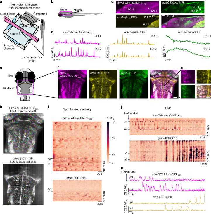
Fig. 4 Three-color multiplexed functional imaging in zebrafish larvae.

a, Light-sheet imaging setup for multiplexed imaging. b, Schematic of side-view zebrafish larvae highlighting the field of view for three-color multiplexed functional imaging of glucose and Ca2+ in muscles and neurons. c, Representative images of WHaloCaMP1a expressed in neurons from the elavl3 promoter, iGlucoSnFR expressed from the actb2 ubiquitous promoter and jRGECO1a expressed in muscle from the acta1a promoter. Scale bar, 50 µm. The experiment was repeated independently three times with similar results. d, Fluorescence ∆F/F0 traces of WHaloCaMP1a669, jRGECO1a and iGlucoSnFR in the ROI outlined in b. A representative experiment from three zebrafish larvae was imaged. e, Schematic of the zebrafish larva’s head indicating the field of view for light-sheet imaging of neuronal and astrocyte activity. f, Representative images of the expression patterns of WHaloCaMP1a669–EGFP expressed under the elavl3 promoter and jRGECO1b expressed under the gfap promoter. Scale bar, 50 µm. The experiment was repeated independently more than three times with similar results. g, Zoomed-in images showing single-cell resolution of fluorescent signals in the hindbrain. Scale bar, 20 µm. h, Images of Suite2p- and Cellpose-segmented cells from simultaneous functional imaging of WHaloCaMP1a669 and jRGECO1b. i, Rastermaps of activity from 1,228 segmented neurons (top) and 530 astrocytes (bottom) during spontaneous brain activity. Two neurons (n1 and n2) indicate the hindbrain oscillator. Two astrocytes (a1 and a1) are also indicated. j, The compound 4-AP was added to the imaging chamber of the zebrafish larva imaged in i, and functional imaging was performed. Concatenation of three imaging blocks of 6.2 min each. k, Fluorescence ∆F/F0 traces of n1 and n2 (top) and a1 and a2 (bottom) after addition of 4-AP.

Acknowledgments
This image is the copyrighted work of the attributed author or publisher, and ZFIN has permission only to display this image to its users. Additional permissions should be obtained from the applicable author or publisher of the image. Full text @ Nat. Methods