IMAGE

Fig. 2

ID
ZDB-IMAGE-241017-46
Source
Figures for Ishizaka et al., 2024
Image
Figure Caption

Fig. 2

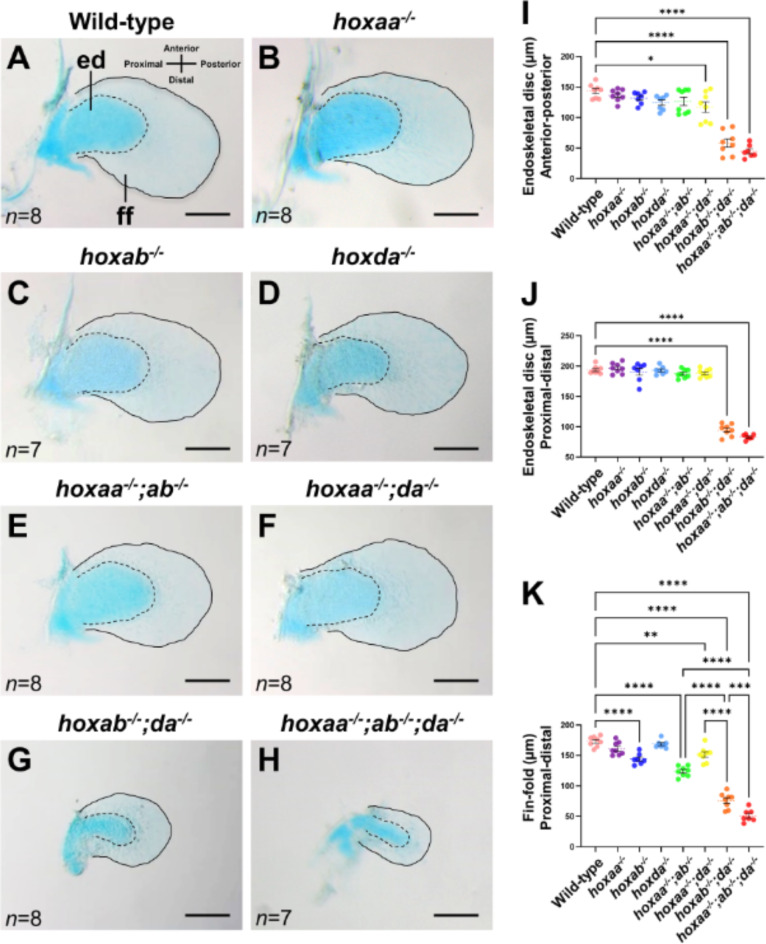
Shortening of the endoskeletal disc and fin-fold of the pectoral fins in hoxaa−/−;hoxab−/−;hoxda−/− larvae. (AH) Reduction of the endoskeletal disc (ed) and fin-fold (ff) in the pectoral fins of hoxaa; hoxab; hoxda cluster-deleted mutants. Alcian blue staining was performed to distinguish cartilaginous cells (endoskeletal disc) and non-cartilaginous fin-fold in larvae at 5 dpf. After the staining, the pectoral fins were manually dissected and mounted on a glass slide. The margin of the endoskeletal disc is emphasized with a dashed line, and the margin of the fin-fold is highlighted with a line. Proximal to the left. Scale bar, 100 μm. The number of dissected pectoral fins for which lengths were measured is noted in the left bottom. (IK) Comparisons of the length of the endoskeletal disc and fin-fold in hoxaa; hoxab; hoxda cluster-deleted mutants. The lengths of the endoskeletal disc and the fin-fold were measured by ImageJ. The Tukey–Kramer test was performed with *P < 0.05, **P < 0.01, ***P < 0.001, and ****P < 0.0001. Error bars represent the standard error of the mean.

Acknowledgments
This image is the copyrighted work of the attributed author or publisher, and ZFIN has permission only to display this image to its users. Additional permissions should be obtained from the applicable author or publisher of the image. Full text @ Sci. Rep.