IMAGE

Fig. 6

ID
ZDB-IMAGE-241017-36
Source
Figures for Armistead et al., 2024
Image
Figure Caption

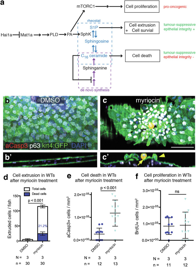
Fig. 6 Early inhibition of ceramide synthesis in wild-type embryos recapitulates ACE and apoptosis phenotype.

a Loss of Matriptase inhibition leads to parallel pro-oncogenic and tumour-suppressive pathways. Sphingolipid rheostat-mediated tumour-suppressive methods include S1P-dependent apical cell extrusion, which preserves epithelial integrity, and C16 ceramide-dependent apoptotic cell death, which may lead to loss of epithelial barrier function. When levels of C16 ceramide are above a certain threshold, a negative feedback loop prevents further de novo ceramide synthesis. In hai1afr26 mutants by 2 dpf, ceramide levels drop due to sustained SphK activity catalysing S1P production, repression of de novo synthesis is lost, and resultant C16 ceramide levels trigger apoptosis. b-c Epidermal aggregates and apoptotic peridermal cells in the caudal fin fold of 4 dpf WT fish, transiently treated with inhibitor of de novo ceramide synthesis myriocin at 2 dpf, with orthogonal views (b’, c’) showing extruding live (yellow arrowhead) or apoptotic (yellow arrow) peridermal cells. Apoptotic cells are labelled with aCasp3 (red), peridermal cells with krt4:GFP (green), basal keratinocytes with p63 (white), and nuclei using DAPI (blue). Scale bar = 50 μm. d Quantification of the numbers of extruded cells collected per fish at 4 dpf upon inhibition of de novo sphingolipid synthesis by myriocin treatment at 2 dpf. White bars show the numbers of live cells, blue bars show the numbers of dead cells, with the proportion of dead cells indicated with blue text. e Quantification of aCasp3-positive cells in the tail fins of 4 dpf embryos upon myriocin treatment, normalised to fin area. f Quantification of proliferating cells in the tail fins of 4 dpf embryos upon myriocin treatment, normalised to fin area. For all graphs, means were compared via two-tailed, unpaired Student’s t-test. N = number of biological replicates, n = number of fish per condition.

Acknowledgments
This image is the copyrighted work of the attributed author or publisher, and ZFIN has permission only to display this image to its users. Additional permissions should be obtained from the applicable author or publisher of the image. Full text @ Cell Death Dis.