
IMAGE
Fig. 6
- ID
- ZDB-IMAGE-241016-6
- Publication
- McCann et al., 2024 - Emc1 is essential for vision and zebrafish photoreceptor outer segment morphogenesis
- All Figures
- Figures for McCann et al., 2024
Image
|
Figure Caption
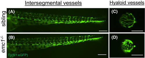
Fig. 6 Hyaloid vessels are dysregulated in emc1−/− zebrafish. (A and B): Fluorescent images displaying lateral views of the trunk of wild-type sibling (A) and emc1−/− (B) larvae at 5 dpf carrying the Tg(fli1:GFP) transgene (green). Scale bar is 200 μm. (C and D): Fluorescent images displaying lateral views of dissected lenses from wild-type siblings (C) and emc1−/− (D) larvae at 5 dpf carrying the Tg(fli1:GFP) transgene (green) to image the hyaloid vessels. Scale bar is 50 μm.
Acknowledgments
This image is the copyrighted work of the attributed author or publisher, and
ZFIN has permission only to display this image to its users.
Additional permissions should be obtained from the applicable author or publisher of the image.
Full text @ FASEB J.